
PIAS1+PIAS2 Rabbit mAb, Unconjugated, Monoclonal
Catalog Number:
ABB-A9670
- Images (8)

| Article Name: | PIAS1+PIAS2 Rabbit mAb, Unconjugated, Monoclonal |
| Biozol Catalog Number: | ABB-A9670 |
| Supplier Catalog Number: | A9670 |
| Alternative Catalog Number: | ABB-A9670-100UL,ABB-A9670-20UL |
| Manufacturer: | ABclonal |
| Host: | Rabbit |
| Category: | Antikörper |
| Application: | ELISA, IHC-P, WB |
| Species Reactivity: | Human |
| Immunogen: | Synthetic peptide. This information is considered to be commercially sensitive. |
| Conjugation: | Unconjugated |
| Alternative Names: | DDXBP1, GBP, GU/RH-II, ZMIZ3, PIAS1+PIAS2 |
| This gene encodes a member of the protein inhibitor of activated STAT (PIAS) family. PIAS proteins function as SUMO E3 ligases and play important roles in many cellular processes by mediating the sumoylation of target proteins. This protein plays a central role as a transcriptional coregulator of numerous cellular pathways includign the STAT1 and nuclear factor kappaB pathways. Alternate splicing results in multiple transcript variants. [provided by RefSeq, Mar 2016] |
| Application Dilute: | WB,1:500 - 1:2000|IHC-P,1:50 - 1:200|ELISA,Recommended starting concentration is 1 µg/mL. Please optimize the concentration based on your specific assay requirements. |
| Application Notes: | Cross-Reactivity: Human,Mouse,Rat. Shipping: Ice Bag |
|
|
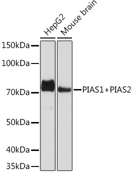
Western blot analysis of various lysates using PIAS1+PIAS2 Rabbit mAb (A9670) at 1:1000 dilution. Secondary antibody: HRP-conjugated Goat anti-Rabbit IgG (H+L) (AS014) at 1:10000 dilution. Lysates/proteins: 25µg per lane. Blocking buffer: 3% nonfat dry milk in TBST. Detection: ECL Basic Kit (RM00020). Exposure time: 90s. |
|
|
Immunohistochemistry analysis of paraffin-embeddedHuman thyroid tissue usingPIAS1+PIAS2 Rabbit mAb(A9670) at a dilution of 1:200 (40x lens).High pressure antigen retrieval was performed with 0.01 M Tris-EDTA buffer (pH 9.0) prior to IHC staining. |
|
|
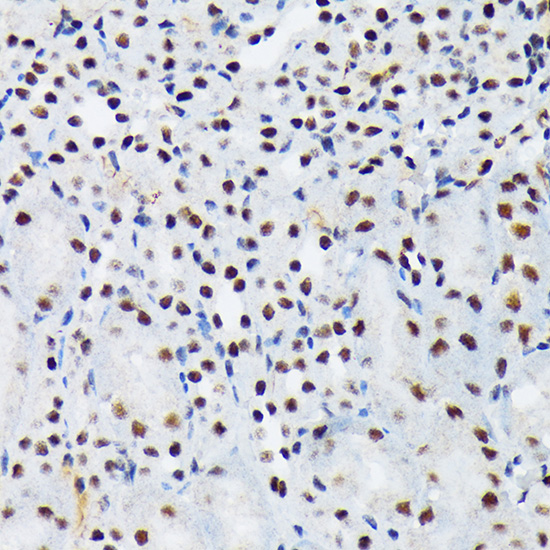
Immunohistochemistry analysis of paraffin-embeddedHuman liver tissue usingPIAS1+PIAS2 Rabbit mAb(A9670) at a dilution of 1:200 (40x lens).High pressure antigen retrieval was performed with 0.01 M Tris-EDTA buffer (pH 9.0) prior to IHC staining. |
|
|
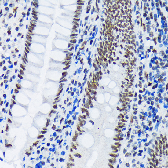
Immunohistochemistry analysis of paraffin-embeddedMouse colon tissue usingPIAS1+PIAS2 Rabbit mAb(A9670) at a dilution of 1:200 (40x lens).High pressure antigen retrieval was performed with 0.01 M Tris-EDTA buffer (pH 9.0) prior to IHC staining. |
|
|
Immunohistochemistry analysis of paraffin-embeddedMouse heart tissue usingPIAS1+PIAS2 Rabbit mAb(A9670) at a dilution of 1:200 (40x lens).High pressure antigen retrieval was performed with 0.01 M Tris-EDTA buffer (pH 9.0) prior to IHC staining. |
|
|
Immunohistochemistry analysis of paraffin-embeddedRat colon tissue usingPIAS1+PIAS2 Rabbit mAb(A9670) at a dilution of 1:200 (40x lens).High pressure antigen retrieval was performed with 0.01 M Tris-EDTA buffer (pH 9.0) prior to IHC staining. |
|
|
Immunohistochemistry analysis of paraffin-embeddedHuman colon tissue usingPIAS1+PIAS2 Rabbit mAb(A9670) at a dilution of 1:200 (40x lens).High pressure antigen retrieval was performed with 0.01 M Tris-EDTA buffer (pH 9.0) prior to IHC staining. |
|
|
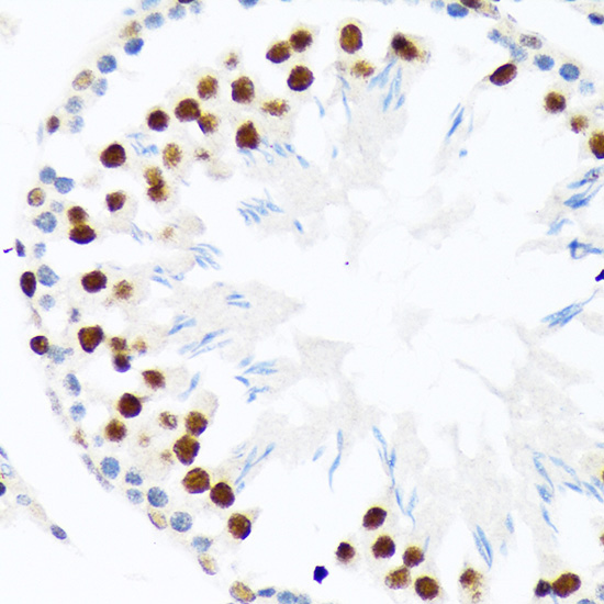
Immunohistochemistry analysis of paraffin-embeddedRat testis tissue usingPIAS1+PIAS2 Rabbit mAb(A9670) at a dilution of 1:200 (40x lens).High pressure antigen retrieval was performed with 0.01 M Tris-EDTA buffer (pH 9.0) prior to IHC staining. |
Product Guarantee and Expert Support
