
Symmetric DiMethyl-Histone H3-R8 Rabbit mAb, Unconjugated
Artikelnummer:
ABB-A21207
- Bilder (8)

| Artikelname: | Symmetric DiMethyl-Histone H3-R8 Rabbit mAb, Unconjugated |
| Artikelnummer: | ABB-A21207 |
| Hersteller Artikelnummer: | A21207 |
| Alternativnummer: | ABB-A21207-20UL,ABB-A21207-100UL,ABB-A21207-1000UL,ABB-A21207-500UL |
| Hersteller: | ABclonal |
| Wirt: | Rabbit |
| Kategorie: | Antikörper |
| Applikation: | DOT, ELISA, IF, IHC-P, WB |
| Spezies Reaktivität: | Human |
| Immunogen: | Synthetic peptide. This information is considered to be commercially sensitive. |
| Konjugation: | Unconjugated |
| Alternative Synonym: | H3/A, H3C2, H3C3, H3C4, H3C6, H3C7, H3C8, H3FA, H3C10, H3C11, H3C12, HIST1H3A, Symmetric DiMethyl-Histone H3-R8 |
| Histones are basic nuclear proteins that are responsible for the nucleosome structure of the chromosomal fiber in eukaryotes. This structure consists of approximately 146 bp of DNA wrapped around a nucleosome, an octamer composed of pairs of each of the four core histones (H2A, H2B, H3, and H4). The chromatin fiber is further compacted through the interaction of a linker histone, H1, with the DNA between the nucleosomes to form higher order chromatin structures. This gene is intronless and encodes a replication-dependent histone that is a member of the histone H3 family. Transcripts from this gene lack polyA tails, instead, they contain a palindromic termination element. This gene is found in the large histone gene cluster on chromosome 6p22-p21.3. |
| Klonalität: | Monoclonal |
| Klon-Bezeichnung: | [ARC53358] |
| Molekulargewicht: | 16kDa |
| NCBI: | 8290 |
| UniProt: | Q16695 |
| Reinheit: | Affinity purification |
| Sequenz: | MARTKQTARKSTGGKAPRKQLATKAARKSAPATGGVKKPHRYRPGTVALREIRRYQKSTELLIRKLPFQRLVREIAQDFKTDLRFQSSAVMALQEACEAY |
| Target-Kategorie: | Histone H3 |
| Antibody Type: | Primary Antibody |
| Application Verdünnung: | WB,1:2000 - 1:10000|DB,1:10000 - 1:120000|IHC-P,1:500 - 1:1000|IF/ICC,1:50 - 1:200|ELISA,Recommended starting concentration is 1 µg/mL. Please optimize the concentration based on your specific assay requirements. |
| Anwendungsbeschreibung: | Cross-Reactivity: Human,Mouse,Rat,Other (Wide Range Predicted). ResearchArea: Protein phosphorylation,Signal Transduction,MAPK-Erk Signaling Pathway,Cell Biology Developmental Biology,Cell Cycle,Immunology Inflammation,NF-kB Signaling Pathway, Epigenetics & Nuclear Signaling,Epigenetic Modifications,Methylation. Shipping: Ice Bag |
|
|
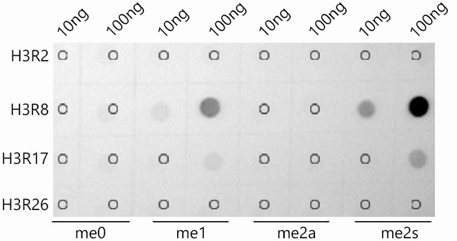
Dot-blot analysis of all sorts of peptides using Symmetric DiMethyl-Histone H3-R8 antibody (A21207) at 1:1000 dilution. |
|
|
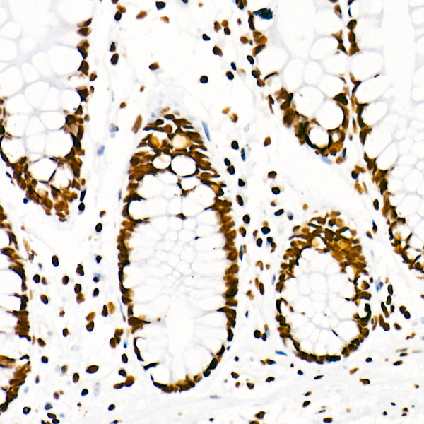
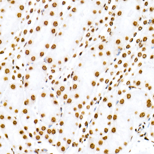
Immunohistochemistry analysis of paraffin-embedded Human colon using Symmetric DiMethyl-Histone H3-R8 Rabbit mAb (A21207) at dilution of 1:1000 (40x lens). High pressure antigen retrieval performed with 0.01M Citrate buffer (pH 6.0) prior to IHC staining. |
|
|
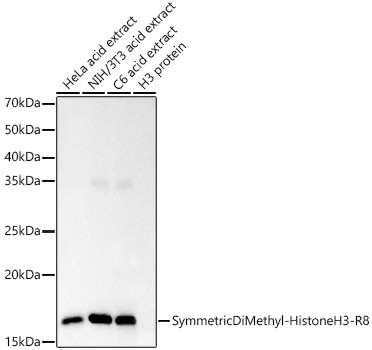
Western blot analysis of various lysates, using Symmetric DiMethyl-Histone H3-R8 Rabbit mAb (A21207) at 1:10000 dilution. Secondary antibody: HRP-conjugated Goat anti-Rabbit IgG (H+L) (AS014) at 1:10000 dilution. Lysates/proteins: 25µg per lane. Blocking buffer: 3% nonfat dry milk in TBST. Detection: ECL Basic Kit (RM00020). Exposure time: 3s. |
|
|
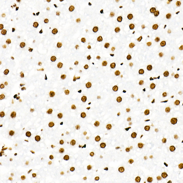
Immunohistochemistry analysis of paraffin-embedded Rat liver using Symmetric DiMethyl-Histone H3-R8 Rabbit mAb (A21207) at dilution of 1:1000 (40x lens). High pressure antigen retrieval performed with 0.01M Citrate buffer (pH 6.0) prior to IHC staining. |
|
|
Immunohistochemistry analysis of paraffin-embedded Mouse kidney using Symmetric DiMethyl-Histone H3-R8 Rabbit mAb (A21207) at dilution of 1:1000 (40x lens). High pressure antigen retrieval performed with 0.01M Citrate buffer (pH 6.0) prior to IHC staining. |
|
|
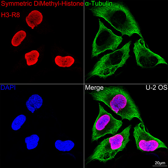
Confocal imaging of PC-12 cells using Symmetric DiMethyl-Histone H3-R8 Rabbit mAb (A21207, dilution 1:200) followed by a further incubation with Cy3-conjugated Goat anti-Rabbit IgG (H+L) (AS007, dilution 1:500) (Red). The cells were counterstained with alpha-Tubulin Mouse mAb (AC012, dilution 1:400) followed by incubation with ABflo 488-conjugated Goat Anti-Mouse IgG (H+L) Ab (AS076, dilution 1:500) (Green). DAPI was used for nuclear staining (Blue). Objective: 100x. |
|
|
Confocal imaging of HeLa cells using Symmetric DiMethyl-Histone H3-R8 Rabbit mAb (A21207,dilution 1:200) followed by a further incubation with Cy3 Goat Anti-Rabbit IgG (H+L) (AS007,dilution 1:500)(Red).The cells were counterstained with alpha-Tubulin Mouse mAb (AC012, dilution 1:400) followed by incubation with ABflo 488-conjugated Goat Anti-Mouse IgG (H+L) Ab (AS076, dilution 1:500) (Green).DAPI was used for nuclear staining (Blue). Objective: 100x. |
|
|
Confocal imaging of NIH/3T3 cells using Symmetric DiMethyl-Histone H3-R8 Rabbit mAb (A21207,dilution 1:200) followed by a further incubation with Cy3 Goat Anti-Rabbit IgG (H+L) (AS007,dilution 1:500)(Red).The cells were counterstained with alpha-Tubulin Mouse mAb (AC012, dilution 1:400) followed by incubation with ABflo 488-conjugated Goat Anti-Mouse IgG (H+L) Ab (AS076, dilution 1:500) (Green).DAPI was used for nuclear staining (Blue). Objective: 100x. |
Produktgarantie und fachkundiger Support
