
RSK2 / RPS6KA3 Rabbit mAb, Unconjugated, Monoclonal
Artikelnummer:
ABB-A20868
- Bilder (8)

| Artikelname: | RSK2 / RPS6KA3 Rabbit mAb, Unconjugated, Monoclonal |
| Artikelnummer: | ABB-A20868 |
| Hersteller Artikelnummer: | A20868 |
| Alternativnummer: | ABB-A20868-100UL,ABB-A20868-20UL |
| Hersteller: | ABclonal |
| Wirt: | Rabbit |
| Kategorie: | Antikörper |
| Applikation: | ELISA, IHC-P, WB |
| Spezies Reaktivität: | Human |
| Immunogen: | Synthetic peptide. This information is considered to be commercially sensitive. |
| Konjugation: | Unconjugated |
| Alternative Synonym: | CLS, RSK, HU-3, RSK2, MRX19, ISPK-1, XLID19, p90-RSK2, pp90RSK2, MAPKAPK1B, S6K-alpha3, RSK2 / RPS6KA3 |
| This gene encodes a member of the RSK (ribosomal S6 kinase) family of serine/threonine kinases. This kinase contains 2 non-identical kinase catalytic domains and phosphorylates various substrates, including members of the mitogen-activated kinase (MAPK) signalling pathway. The activity of this protein has been implicated in controlling cell growth and differentiation. Mutations in this gene have been associated with Coffin-Lowry syndrome (CLS). |
| Application Verdünnung: | WB,1:100 - 1:500|IHC-P,1:50 - 1:200|ELISA,Recommended starting concentration is 1 µg/mL. Please optimize the concentration based on your specific assay requirements. |
| Anwendungsbeschreibung: | Cross-Reactivity: Human,Mouse,Rat. ResearchArea: Epigenetics Nuclear Signaling,Translation Control,Regulation of eIF4 and p70 S6 Kinase,Signal Transduction,G protein signaling,G-Protein-Coupled Receptors Signaling to MAPK Erk,Kinase,Serine threonine kinases,mTOR Signaling Pathway,MAPK-Erk Signaling Pathway,Cell Biology Developmental Biology,Apoptosis,Mitochondrial Control of Apoptosis,Inhibition of Apoptosis,Cell Cycle,Cell Cycle Control-G2 M DNA Damage Checkpoint,Microtubules,Immunology Inflammation,Neuroscience. Shipping: Ice Bag |
|
|
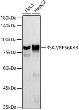
Western blot analysis of various lysates using RSK2 / RPS6KA3 Rabbit mAb (A20868) at 1:500 dilution. Secondary antibody: HRP-conjugated Goat anti-Rabbit IgG (H+L) (AS014) at 1:10000 dilution. Lysates/proteins: 25µg per lane. Blocking buffer: 3% nonfat dry milk in TBST. Detection: ECL Basic Kit (RM00020). Exposure time: 60s. |
|
|
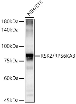
Western blot analysis of lysates from NIH/3T3 cells, using RSK2 / RPS6KA3 Rabbit mAb (A20868) at 1:500 dilution. Secondary antibody: HRP-conjugated Goat anti-Rabbit IgG (H+L) (AS014) at 1:10000 dilution. Lysates/proteins: 25µg per lane. Blocking buffer: 3% nonfat dry milk in TBST. Detection: ECL Basic Kit (RM00020). Exposure time: 180s. |
|
|
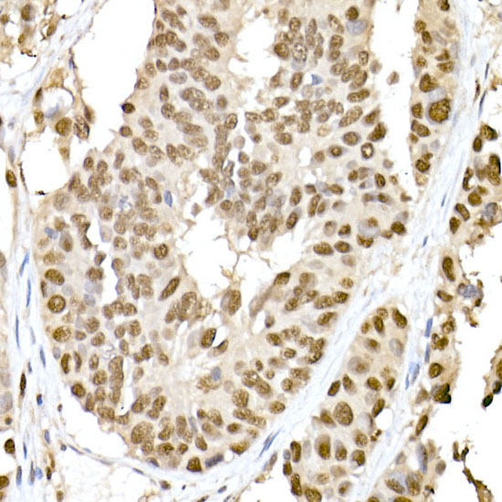
Immunohistochemistry analysis of paraffin-embeddedHuman breast cancer tissue usingRSK2 / RPS6KA3 Rabbit mAb(A20868) at a dilution of 1:200 (40x lens).High pressure antigen retrieval was performed with 0.01 M citrate buffer (pH 6.0) prior to IHC staining. |
|
|
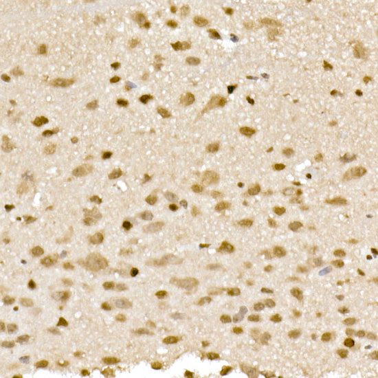
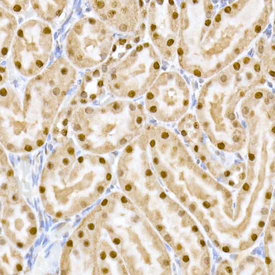
Immunohistochemistry analysis of paraffin-embeddedHuman liver tissue usingRSK2 / RPS6KA3 Rabbit mAb(A20868) at a dilution of 1:200 (40x lens).High pressure antigen retrieval was performed with 0.01 M citrate buffer (pH 6.0) prior to IHC staining. |
|
|
Immunohistochemistry analysis of paraffin-embeddedRat intestine tissue usingRSK2 / RPS6KA3 Rabbit mAb(A20868) at a dilution of 1:200 (40x lens).High pressure antigen retrieval was performed with 0.01 M citrate buffer (pH 6.0) prior to IHC staining. |
|
|
Immunohistochemistry analysis of paraffin-embeddedMouse lung tissue usingRSK2 / RPS6KA3 Rabbit mAb(A20868) at a dilution of 1:200 (40x lens).High pressure antigen retrieval was performed with 0.01 M citrate buffer (pH 6.0) prior to IHC staining. |
|
|
Immunohistochemistry analysis of paraffin-embeddedMouse intestin tissue usingRSK2 / RPS6KA3 Rabbit mAb(A20868) at a dilution of 1:200 (40x lens).High pressure antigen retrieval was performed with 0.01 M citrate buffer (pH 6.0) prior to IHC staining. |
|
|
Immunohistochemistry analysis of paraffin-embeddedRat liver tissue usingRSK2 / RPS6KA3 Rabbit mAb(A20868) at a dilution of 1:200 (40x lens).High pressure antigen retrieval was performed with 0.01 M citrate buffer (pH 6.0) prior to IHC staining. |
Produktgarantie und fachkundiger Support
