
RAB5A Rabbit pAb, Unconjugated, Polyclonal
Artikelnummer:
ABB-A1180
- Bilder (8)


| Artikelname: | RAB5A Rabbit pAb, Unconjugated, Polyclonal |
| Artikelnummer: | ABB-A1180 |
| Hersteller Artikelnummer: | A1180 |
| Alternativnummer: | ABB-A1180-20UL,ABB-A1180-100UL |
| Hersteller: | ABclonal |
| Wirt: | Rabbit |
| Kategorie: | Antikörper |
| Applikation: | ELISA, IF, IHC-P, WB |
| Spezies Reaktivität: | Human |
| Immunogen: | This information is considered to be commercially sensitive. |
| Konjugation: | Unconjugated |
| Alternative Synonym: | RAB5, RAB5A |
| Enables GDP binding activity, GTP binding activity, and GTPase activity. Involved in several processes, including amyloid-beta clearance by transcytosis, early endosome to late endosome transport, and regulation of exocytosis. Located in several cellular components, including cytoplasmic side of early endosome membrane, nucleoplasm, and terminal bouton. |
| Application Verdünnung: | WB,1:500 - 1:1000|IHC-P,1:50 - 1:200|IF/ICC,1:50 - 1:200|ELISA,Recommended starting concentration is 1 µg/mL. Please optimize the concentration based on your specific assay requirements. |
| Anwendungsbeschreibung: | Cross-Reactivity: Human,Mouse,Rat. ResearchArea: Signal Transduction,Neuroscience, Cell Type Marker,Neurodegenerative Diseases,Neuron marker,Synapse marker. Shipping: Ice Bag |
|
|
Western blot analysis of various lysates, using RAB5A Rabbit pAb (A1180) at 1:700 dilution. Secondary antibody: HRP-conjugated Goat anti-Rabbit IgG (H+L) (AS014) at 1:10000 dilution. Lysates/proteins: 25µg per lane. Blocking buffer: 3% nonfat dry milk in TBST. Detection: ECL Basic Kit (RM00020). Exposure time: 30s. |
|
|
Immunohistochemistry analysis of paraffin-embedded Human liver cancer using RAB5A Rabbit pAb (A1180) at dilution of 1:100 (40x lens). High pressure antigen retrieval performed with 0.01M Citrate buffer (pH 6.0) prior to IHC staining. |
|
|
Immunohistochemistry analysis of paraffin-embedded Human liver using RAB5A Rabbit pAb (A1180) at dilution of 1:100 (40x lens). High pressure antigen retrieval performed with 0.01M Citrate buffer (pH 6.0) prior to IHC staining. |
|
|
Immunohistochemistry analysis of paraffin-embedded Mouse spleen using RAB5A Rabbit pAb (A1180) at dilution of 1:100 (40x lens). High pressure antigen retrieval performed with 0.01M Citrate buffer (pH 6.0) prior to IHC staining. |
|
|
Immunohistochemistry analysis of paraffin-embedded Rat spleen using RAB5A Rabbit pAb (A1180) at dilution of 1:100 (40x lens). High pressure antigen retrieval performed with 0.01M Citrate buffer (pH 6.0) prior to IHC staining. |
|
|
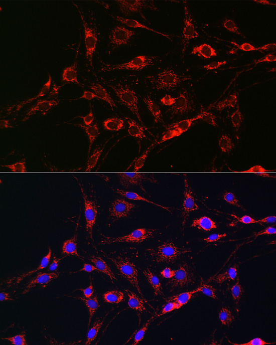
Immunofluorescence analysis of C6 cells using RAB5A Rabbit pAb (A1180) at dilution of 1:100 (40x lens). Secondary antibody: Cy3-conjugated Goat anti-Rabbit IgG (H+L) (AS007) at 1:500 dilution. Blue: DAPI for nuclear staining. |
|
|
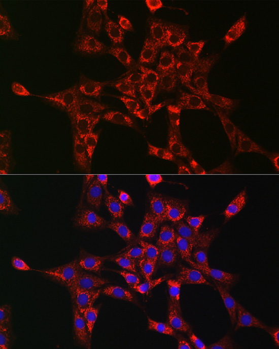
Immunofluorescence analysis of NIH-3T3 cells using RAB5A Rabbit pAb (A1180) at dilution of 1:100 (40x lens). Secondary antibody: Cy3-conjugated Goat anti-Rabbit IgG (H+L) (AS007) at 1:500 dilution. Blue: DAPI for nuclear staining. |
|
|
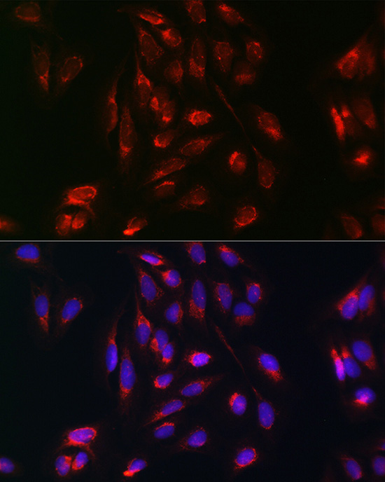
Immunofluorescence analysis of U-2 OS cells using RAB5A Rabbit pAb (A1180) at dilution of 1:100 (40x lens). Secondary antibody: Cy3-conjugated Goat anti-Rabbit IgG (H+L) (AS007) at 1:500 dilution. Blue: DAPI for nuclear staining. |
Produktgarantie und fachkundiger Support
