VISTA Antibody is supplied in PBS containing 0.02% sodium azide and 50% glycerol.
Form:
Liquid
Application Dilute:
Optimal dilutions for each application to be determined by the researcher.
Application Notes:
VISTA antibody can be used for detection of VISTA by Western blot at 0.25 - 1 µg/mL. Antibody can also be used for immunohistochemistry starting at 2 µg/mL and immunocytochemistry starting at 1 µg/mL. For immunofluorescence start at 2 µg/mL.Antibody validated: Western Blot in human samples, Immunohistochemistry in human samples, Immunocytochemistry in human samples, Immunofluorescence in human samples and Flow Cytometry in mouse samples. All other applications and species not yet tested.
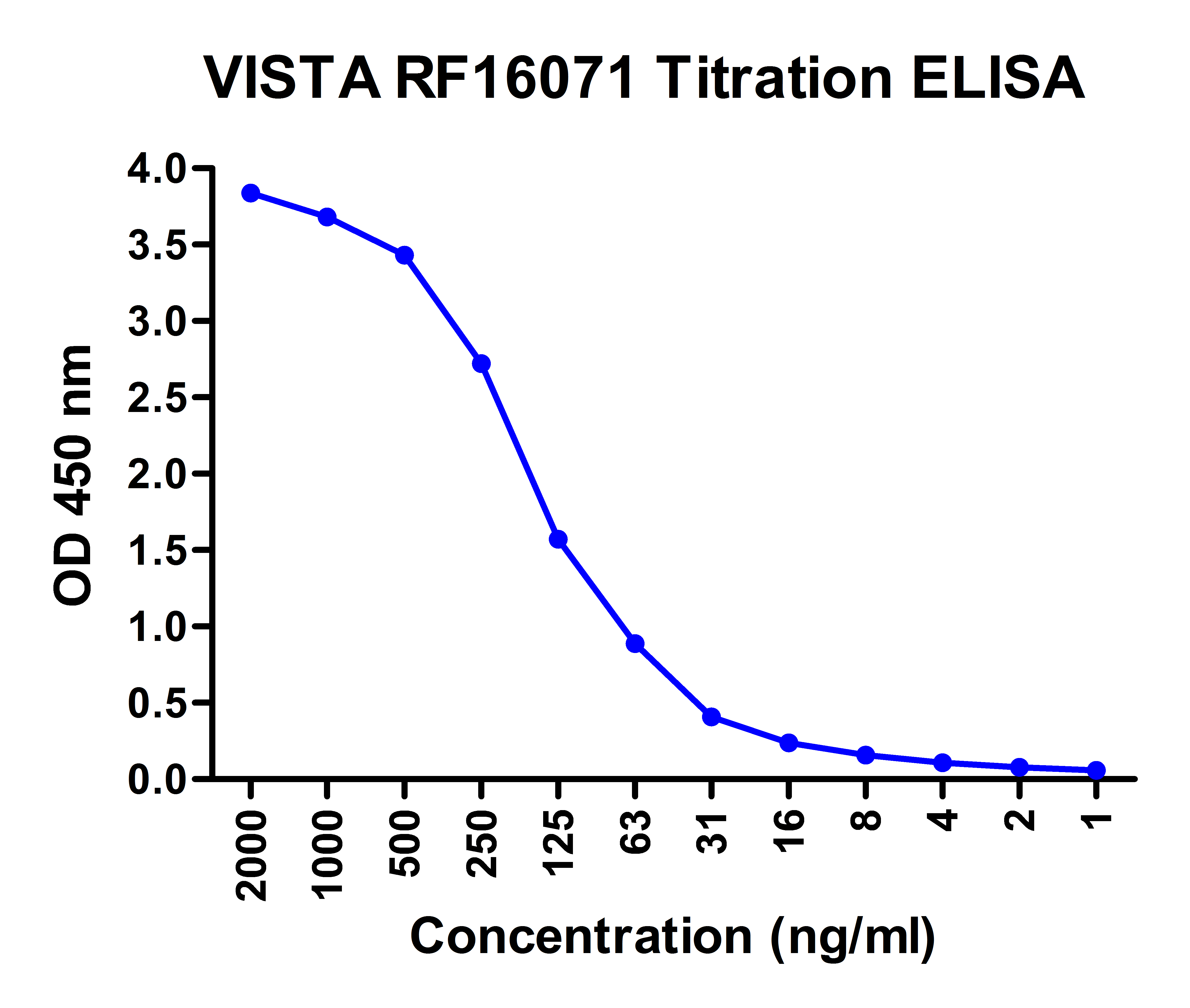
Titration curve analysis of VISTA antibody to detect recombinant VISTA in ELISA at decreasing concentrations.
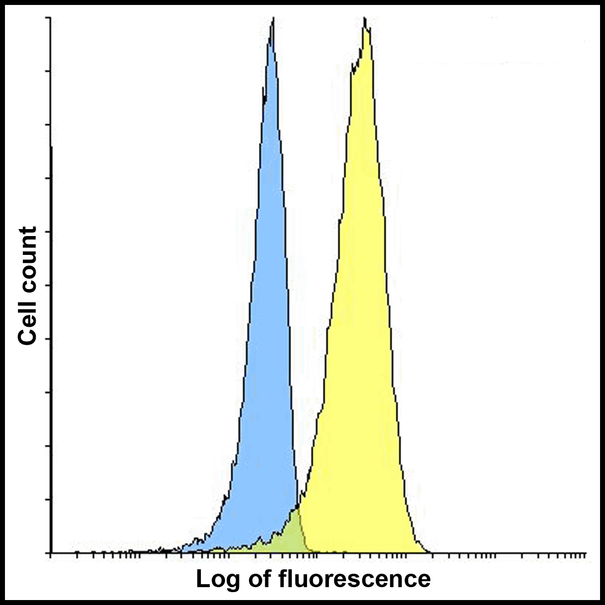
Flow cytometry analysis of VISTA overexpressing HEK293 cells using VISTA antibody and control mouse IgG antibody at 10 &956,g/ml. Blue: Untransfected HEK293 cells. Yellow: VISTA overexpressing HEK293 cells.
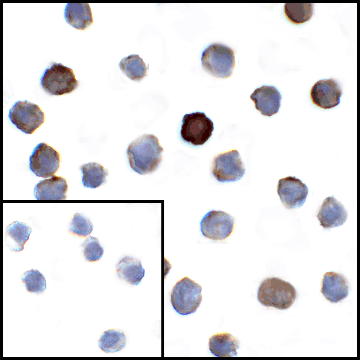
Immunocytochemistry of VISTA in transfected HEK293 cells with VISTA antibody at 1 &956,g/mL. Lower left: Immunocytochemistry in transfected HEK293 cells with control mouse IgG antibody at 1 &956,g/mL.
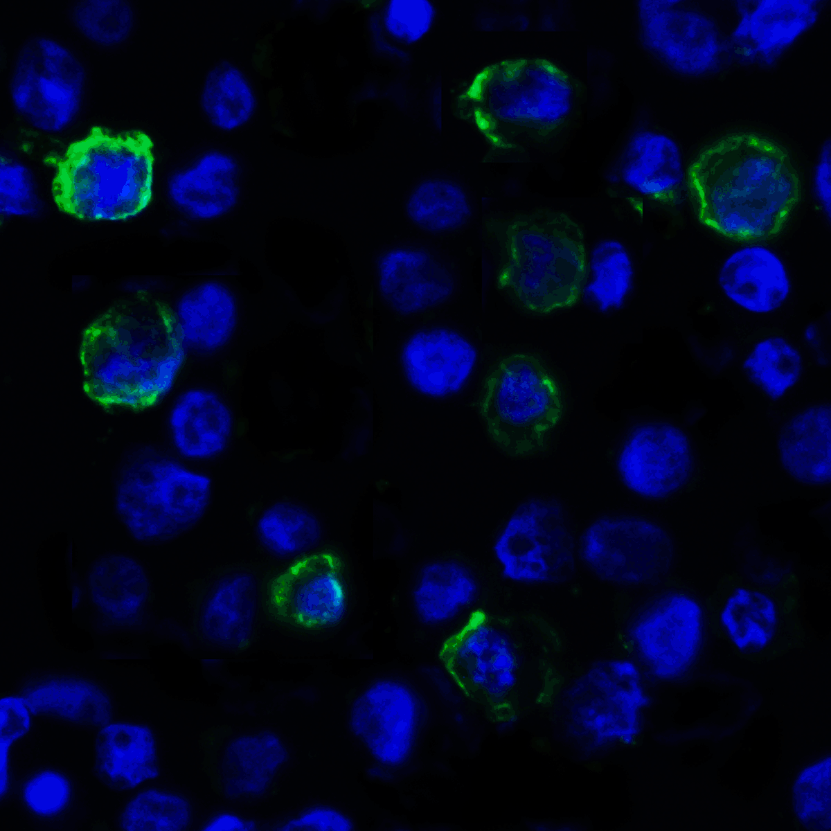
Immunofluorescence of VISTA in transfected HEK293 cells with VISTA antibody at 2 &956,g/mL. Green: VISTA Antibody [4C4] (RF16071) Blue: DAPI staining
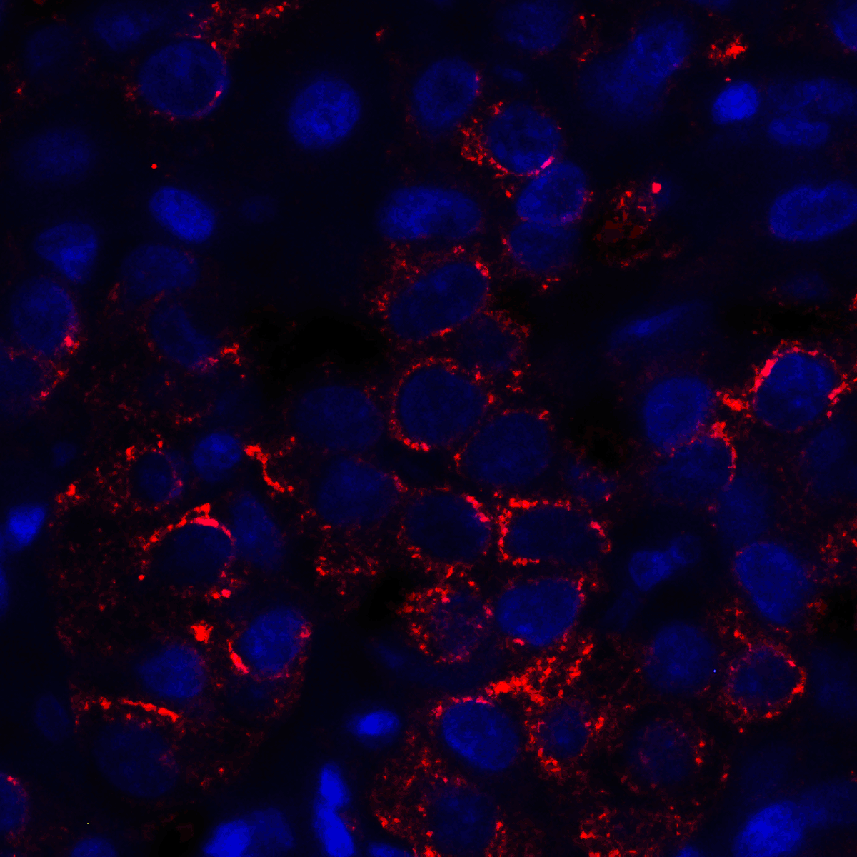
Immunofluorescence of VISTA in human lymphoma tissue with VISTA antibody at 5 &956,g/mL. Red: VISTA Antibody [4C4] (RF16071) Blue: DAPI staining
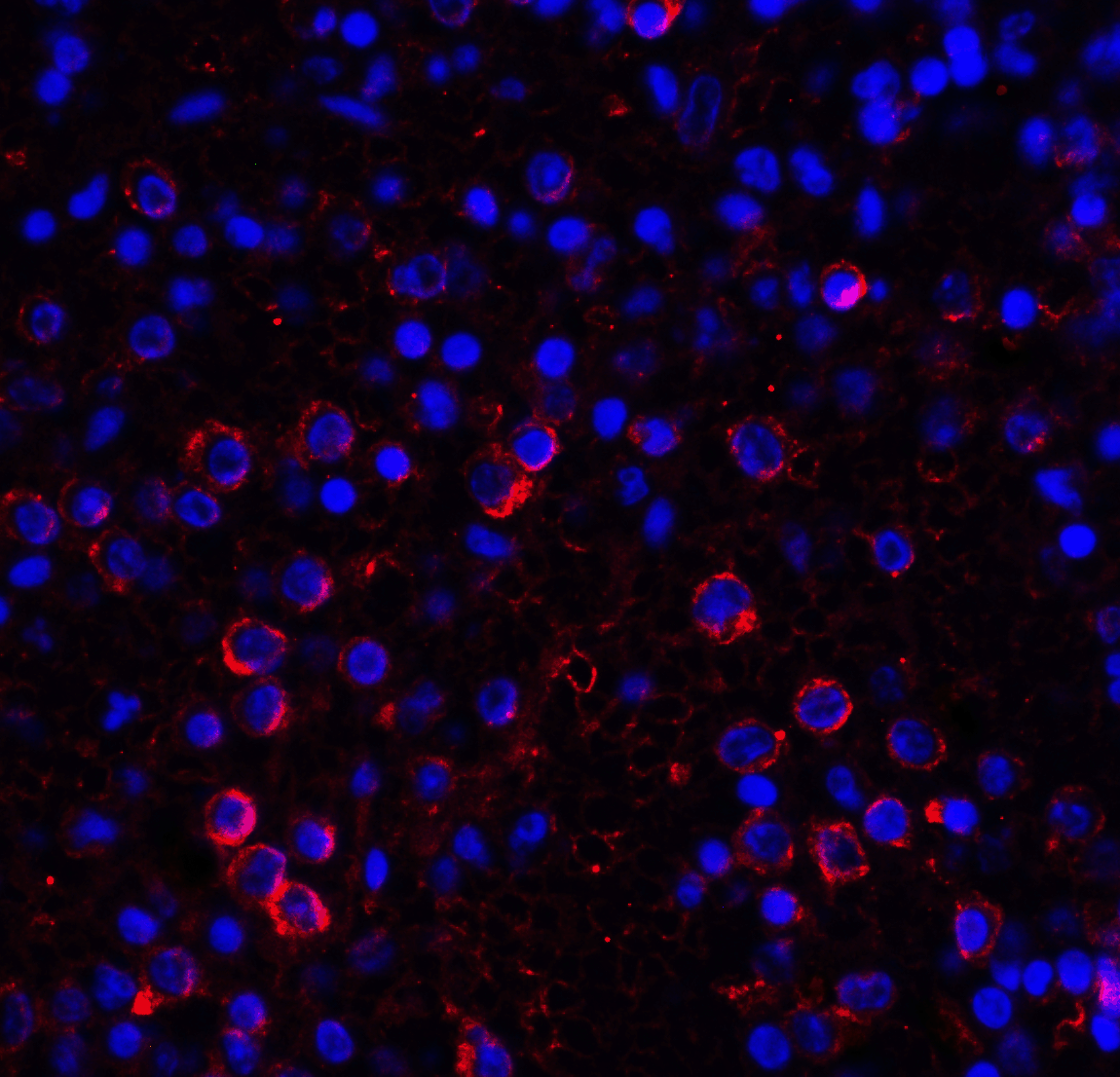
Immunofluorescence of VISTA in human spleen tissue with VISTA antibody at 10 &956,g/mL. Red: VISTA Antibody [4C4] (RF16071) Blue: DAPI staining
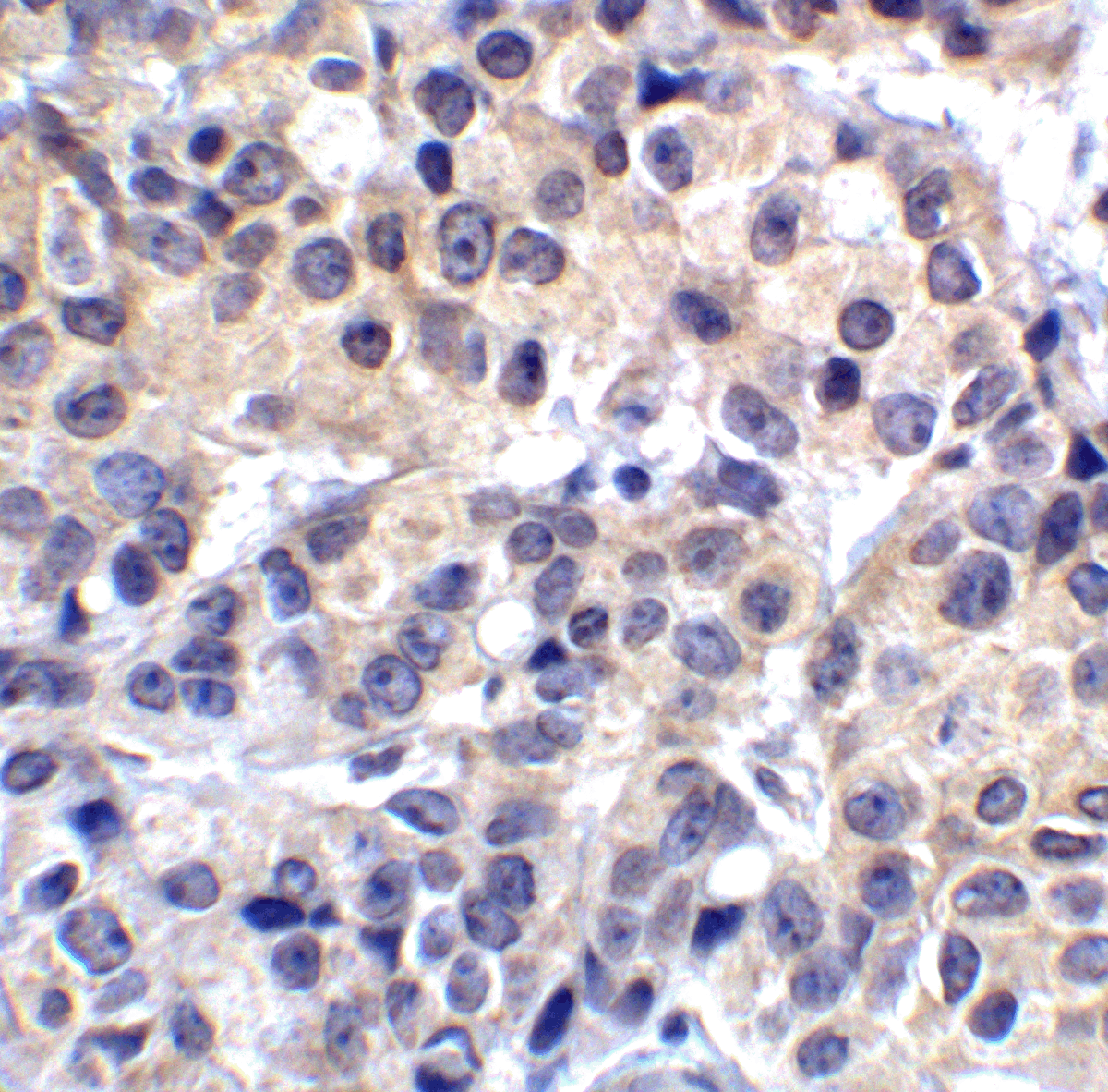
Immunohistochemistry of VISTA in human lymphoma tissue with VISTA antibody at 2 &956,g/mL.
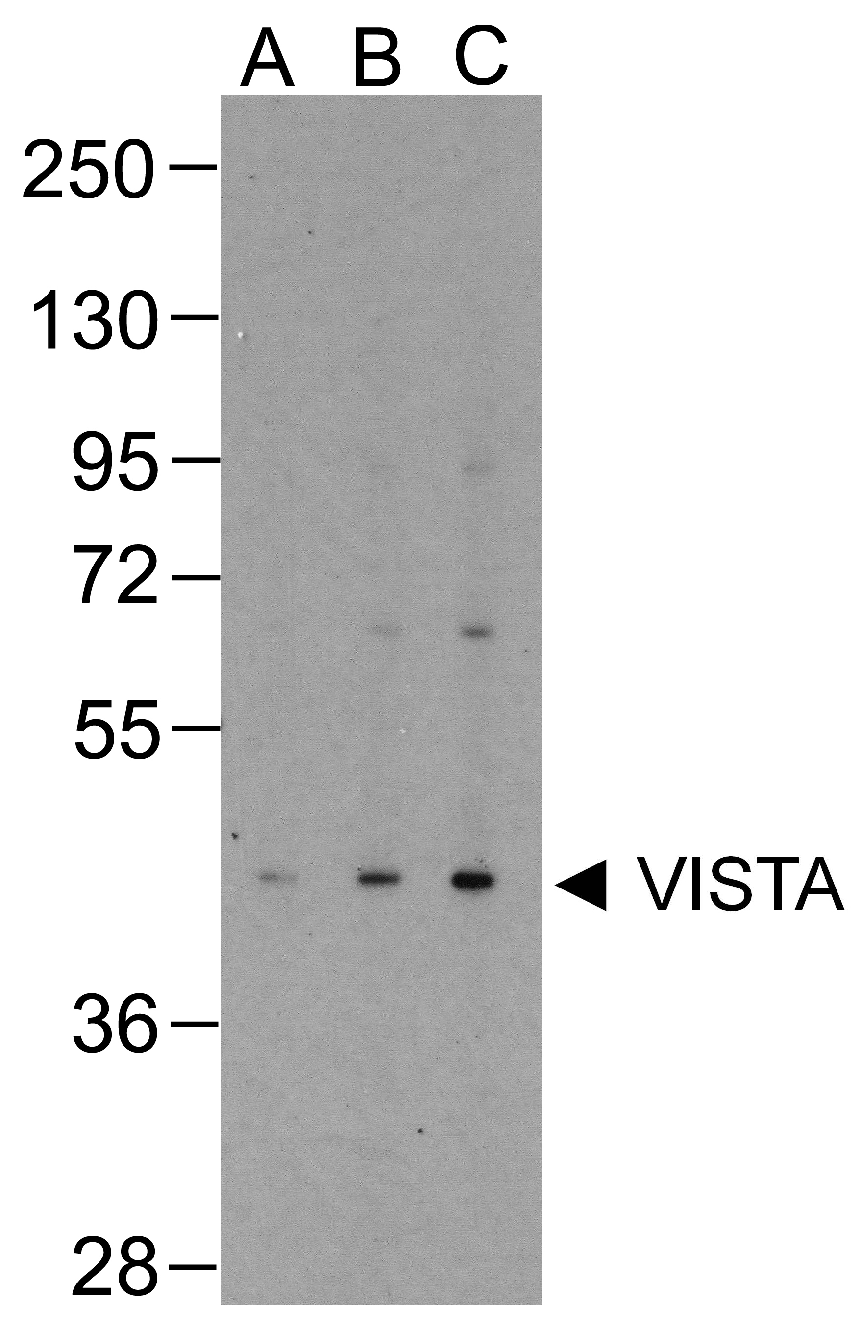
Western blot analysis of VISTA in overexpressing HEK293 cells with VISTA antibody at (A) 0.25 (B) 0.5 and (C) 1 &956,g/ml
* VAT and and shipping costs not included. Errors and price changes excepted