
MANF Antibody, Unconjugated, Rabbit, Polyclonal
Catalog Number:
PRS-4349
- Images (6)


| Article Name: | MANF Antibody, Unconjugated, Rabbit, Polyclonal |
| Biozol Catalog Number: | PRS-4349 |
| Supplier Catalog Number: | 4349 |
| Alternative Catalog Number: | PRS-4349-0.02,PRS-4349-0.1 |
| Manufacturer: | ProSci |
| Host: | Rabbit |
| Category: | Antikörper |
| Application: | ELISA, IF, IHC-P, WB |
| Species Reactivity: | Human, Mouse, Rat |
| Immunogen: | Anti-MANF antibody (4349) was raised against a peptide corresponding to 12 amino acids near the amino terminus of human MANF. The immunogen is located within the first 50 amino acids of MANF. |
| Conjugation: | Unconjugated |
| Alternative Names: | MANF Antibody: ARP, ARMET, ARP, Mesencephalic astrocyte-derived neurotrophic factor, Arginine-rich protein |
| Application Dilute: | Optimal dilutions for each application to be determined by the researcher. |
| Application Notes: | WB: 0.125-2 µg/mL, IHC: 2.5 µg/mL, IF: 20 µg/mL.Antibody validated: Western Blot in human and rat samples, Immunohistochemistry in human samples, Immunofluorescence in human samples. All other applications and species not yet tested. |
|
|
Figure 3 Western Blot Validation in Rat Brain Tissue LysateLoading: 15 &956,g of lysates per lane.Antibodies: MANF 4349 (A: 1 &956,g /mL and B: 2 &956,g /mL), 1h incubation at RT in 5% NFDM/TBST.Secondary: Goat anti-rabbit IgG HRP conjugate at 1:10000 dilution. |
|
|
Figure 5 Immunofluorescence Validation of MANF in Human Brain TissueImmunofluorescent analysis of 4% paraformaldehyde-fixed human brain tissue labeling MANF with 4349 at 20 &956,g /mL, followed by goat anti-rabbit IgG secondary antibody at 1/500 dilution (red). |
|
|
Figure 4 Immunohistochemistry Validation of MANF in Human Brain Tissue Immunohistochemical analysis of paraffin-embedded human brain tissue using anti-MANF antibody (4349) at 2.5 &956,g /ml. Tissue was fixed with formaldehyde and blocked with 10% serum for 1 h at RT, antigen retrieval was by heat mediation with a citrate buffer (pH6). Samples were incubated with primary antibody overnight at 4&730,C. A goat anti-rabbit IgG H&L (HRP) at 1/250 was used as secondary. Counter stained with Hematoxylin. |
|
|
Figure 1 Western Blot Validation in Human Cell LinesLoading: 15 &956,g of lysates per lane.Antibodies: MANF 4349, (2 &956,g/mL), 1h incubation at RT in 5% NFDM/TBST.Secondary: Goat anti-rabbit IgG HRP conjugate at 1:10000 dilution. |
|
|
Figure 2 Western Blot Validation with Recombinant ProteinLoading: 30 ng of human MANF recombinant protein per lane.Antibodies: MANF 4349 (Lane 1: 0.125 &956,g/mL, Lane 2: 0.25 &956,g/mL and Lane 3: 0.5 &956,g/mL), 1h incubation at RT in 5% NFDM/TBST.Secondary: Goat anti-rabbit IgG HRP conjugate at 1:10000 dilution.Observed at around 55kD. |
|
|
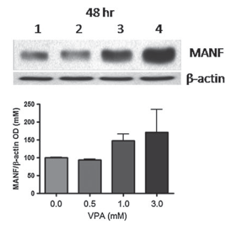
Figure 6 Induced Expression Validation of MANF in Mouse C17.2 Neural Stem Cells (Almutawaa et al., 2014) Concentration- and time-dependent effects of Valproic acid (VPA) on MANF at 48 h examined by Western blot analysis with anti-MANF antibodies. MANF was markedly increased 48h after VPA treatment.Lanes 1-4: control, 0.5 mM VPA, 1 mM VPA, 3 mM VPA. |
Product Guarantee and Expert Support
