
RORC Antibody, Unconjugated, Rabbit, Polyclonal
Catalog Number:
CSB-PA020071LA01HU
- Images (5)


| Article Name: | RORC Antibody, Unconjugated, Rabbit, Polyclonal |
| Biozol Catalog Number: | CSB-PA020071LA01HU |
| Supplier Catalog Number: | CSB-PA020071LA01HU |
| Alternative Catalog Number: | CSB-PA020071LA01HU-100UG, CSB-PA020071LA01HU-50UG |
| Manufacturer: | Cusabio |
| Host: | Rabbit |
| Category: | Antikörper |
| Application: | ELISA, IHC, WB |
| Species Reactivity: | Human |
| Conjugation: | Unconjugated |
| Alternative Names: | IMD42 antibody, MGC129539 antibody, NR1F3 antibody, Nuclear receptor ROR gamma antibody, Nuclear receptor ROR-gamma antibody, Nuclear receptor RZR gamma antibody, Nuclear receptor RZR-gamma antibody, Nuclear receptor subfamily 1 group F member 3 antibody, RAR related orphan nuclear receptor variant 2 antibody, RAR related orphan receptor C antibody, RAR related orphan receptor C, isoform a antibody, RAR related orphan receptor gamma antibody, RAR-related orphan receptor C antibody, Retinoic acid binding receptor gamma antibody, Retinoid related orphan receptor gamma antibody, Retinoid-related orphan receptor-gamma antibody, Rorc antibody, RORG antibody, RORG_HUMAN antibody, RZR GAMMA antibody, RZRG antibody, TOR antibody |
| Clonality: | Polyclonal |
| UniProt: | P51449 |
| Buffer: | Preservative: 0.03% Proclin 300<br />Constituents: 50% Glycerol, 0.01M PBS, PH 7.4 |
| Purity: | >95%, Protein G purified |
| Form: | Liquid |
| Target: | RORC |
| Application Dilute: | Recommended dilution: WB:1:1000-1:5000, IHC:1:20-1:200 |
|
|
IHC image of CSB-PA020071LA01HU diluted at 1:1000 and staining in paraffin-embedded human testis tissue performed on a Leica BondTM system. After dewaxing and hydration, antigen retrieval was mediated by high pressure in a citrate buffer (pH 6.0). Section was blocked with 10% normal goat serum 30min at RT. Then primary antibody (1% BSA) was incubated at 4C overnight. The primary is detected by a Goat anti-rabbit polymer IgG labeled by HRP and visualized using 0.05% DAB.Secondary antibody only control: uses 1% BSA instead of primary antibody |
|
|
IHC image of CSB-PA020071LA01HU diluted at 1:1000 and staining in paraffin-embedded human skin tissue performed on a Leica BondTM system. After dewaxing and hydration, antigen retrieval was mediated by high pressure in a citrate buffer (pH 6.0). Section was blocked with 10% normal goat serum 30min at RT. Then primary antibody (1% BSA) was incubated at 4C overnight. The primary is detected by a Goat anti-rabbit polymer IgG labeled by HRP and visualized using 0.05% DAB. |
|
|
IHC image of CSB-PA020071LA01HU diluted at 1:1000 and staining in paraffin-embedded mouse liver tissue performed on a Leica BondTM system. After dewaxing and hydration, antigen retrieval was mediated by high pressure in a citrate buffer (pH 6.0). Section was blocked with 10% normal goat serum 30min at RT. Then primary antibody (1% BSA) was incubated at 4C overnight. The primary is detected by a Goat anti-rabbit polymer IgG labeled by HRP and visualized using 0.05% DAB. |
|
|
Western Blot Positive WB detected in: HepG2 whole cell lysate All lanes: RORC antibody at 5.2µg/ml Secondary Goat polyclonal to rabbit IgG at 1/50000 dilution Predicted band size: 59, 56 kDa Observed band size: 59 kDa |
|
|
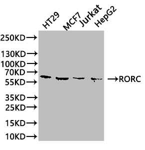
Western Blot Positive WB detected in: HT29 whole cell lysate, MCF7 whole cell lysate, Jurkat whole cell lysate, HepG2 whole cell lysate All lanes: RORC antibody at 1:1000 Secondary Goat polyclonal to rabbit IgG at 1/50000 dilution Predicted band size: 59, 56 kDa Observed band size: 59 kDa |
Product Guarantee and Expert Support
