
PAM Antibody, Unconjugated, Rabbit, Polyclonal
Catalog Number:
CSB-PA017417LA01HU
- Images (4)
| Article Name: | PAM Antibody, Unconjugated, Rabbit, Polyclonal |
| Biozol Catalog Number: | CSB-PA017417LA01HU |
| Supplier Catalog Number: | CSB-PA017417LA01HU |
| Alternative Catalog Number: | CSB-PA017417LA01HU-100UG, CSB-PA017417LA01HU-50UG |
| Manufacturer: | Cusabio |
| Host: | Rabbit |
| Category: | Antikörper |
| Application: | ELISA, IF, IHC, WB |
| Species Reactivity: | Human |
| Conjugation: | Unconjugated |
| Alternative Names: | AMD_HUMAN antibody, PAL antibody, PAM antibody, Pancreatic peptidylglycine alpha amidating monooxygenase antibody, Peptidyl alpha amidating enzyme antibody, Peptidyl alpha hydroxyglycine alpha amidating lyase antibody, Peptidyl-alpha-hydroxyglycine alpha-amidating lyase antibody, Peptidylamidoglycolate lyase antibody, Peptidylglycine 2 hydroxylase antibody, Peptidylglycine alpha amidating monooxygenase antibody, Peptidylglycine alpha hydroxylating monooxygenase antibody, PHM antibody |
| Clonality: | Polyclonal |
| UniProt: | P19021 |
| Buffer: | Preservative: 0.03% Proclin 300<br />Constituents: 50% Glycerol, 0.01M PBS, pH 7.4 |
| Purity: | >95%, Protein G purified |
| Form: | Liquid |
| Target: | PAM |
| Application Dilute: | Recommended dilution: WB:1:500-1:5000, IHC:1:200-1:500, IF:1:50-1:200 |
|
|
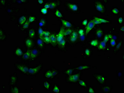
Immunofluorescence staining of Hela cells with CSB-PA017417LA01HU at 1:66, counter-stained with DAPI. The cells were fixed in 4% formaldehyde, permeabilized using 0.2% Triton X-100 and blocked in 10% normal Goat Serum. The cells were then incubated with the antibody overnight at 4°,C. The secondary antibody was Alexa Fluor 488-congugated AffiniPure Goat Anti-Rabbit IgG(H+L). |
|
|
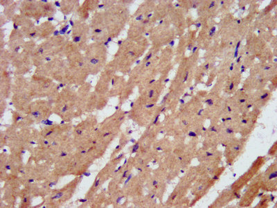
IHC image of CSB-PA017417LA01HU diluted at 1:200 and staining in paraffin-embedded human heart tissue performed on a Leica BondTM system. After dewaxing and hydration, antigen retrieval was mediated by high pressure in a citrate buffer (pH 6.0). Section was blocked with 10% normal goat serum 30min at RT. Then primary antibody (1% BSA) was incubated at 4°,C overnight. The primary is detected by a biotinylated secondary antibody and visualized using an HRP conjugated SP system. |
|
|
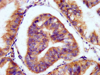
IHC image of CSB-PA017417LA01HU diluted at 1:200 and staining in paraffin-embedded human endometrial cancer performed on a Leica BondTM system. After dewaxing and hydration, antigen retrieval was mediated by high pressure in a citrate buffer (pH 6.0). Section was blocked with 10% normal goat serum 30min at RT. Then primary antibody (1% BSA) was incubated at 4°,C overnight. The primary is detected by a biotinylated secondary antibody and visualized using an HRP conjugated SP system. |
|
|
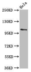
Western Blot Positive WB detected in: Hela whole cell lysate All lanes: PAM antibody at 5.6µg/ml Secondary Goat polyclonal to rabbit IgG at 1/50000 dilution Predicted band size: 109, 97, 101, 99, 107 kDa Observed band size: 109 kDa |
Product Guarantee and Expert Support
