
NPHS1 Antibody, Unconjugated, Rabbit, Polyclonal
Catalog Number:
CSB-PA015988LA01HU
- Images (5)


| Article Name: | NPHS1 Antibody, Unconjugated, Rabbit, Polyclonal |
| Biozol Catalog Number: | CSB-PA015988LA01HU |
| Supplier Catalog Number: | CSB-PA015988LA01HU |
| Alternative Catalog Number: | CSB-PA015988LA01HU-100UG, CSB-PA015988LA01HU-50UG |
| Manufacturer: | Cusabio |
| Host: | Rabbit |
| Category: | Antikörper |
| Application: | ELISA, IHC, WB |
| Species Reactivity: | Human |
| Conjugation: | Unconjugated |
| Alternative Names: | CNF antibody, Nephrin antibody, Nephrosis 1 congenital Finnish type antibody, Nephrosis 1, congenital, Finnish type (nephrin) antibody, NPHN antibody, NPHN_HUMAN antibody, NPHS 1 antibody, Nphs1 antibody, Renal glomerulus specific cell adhesion receptor antibody, Renal glomerulus-specific cell adhesion receptor antibody |
| Clonality: | Polyclonal |
| UniProt: | O60500 |
| Buffer: | Liquid in PBS containing 50% glycerol, 0.5% BSA and 0.02% sodium azide. |
| Purity: | Antigen Affinity Purified |
| Form: | Liquid |
| Target: | NPHS1 |
| Application Dilute: | Recommended dilution: WB:1:1000-1:3000, IHC:1:100-1:300 |
|
|
|
|
|
|
|
|
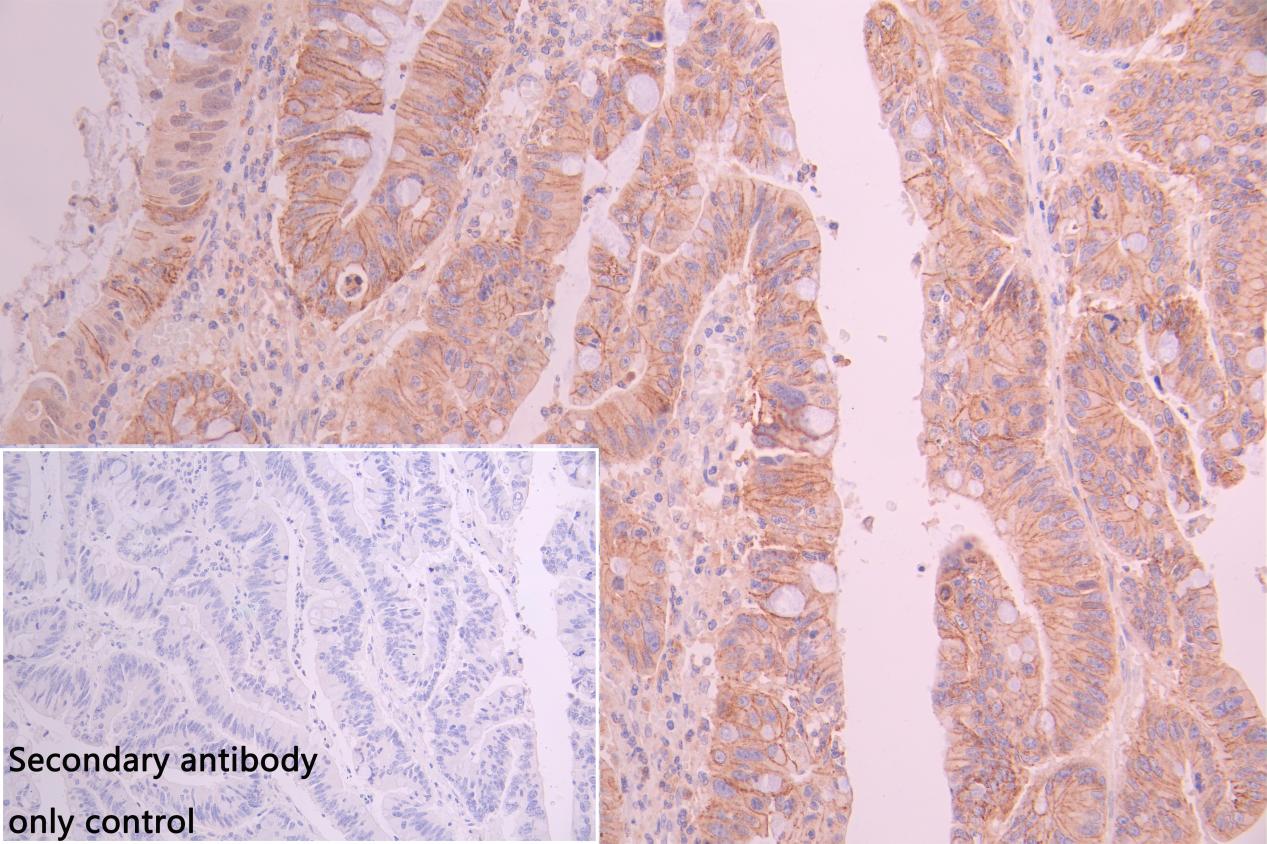
IHC image of CSB-PA015988LA01HU diluted at 1:100 and staining in paraffin-embedded human colorectal cancer performed on a Leica BondTM system. After dewaxing and hydration, antigen retrieval was mediated by high pressure in a citrate buffer (pH 6.0). Section was blocked with 10% normal goat serum 30min at RT. Then primary antibody (1% BSA) was incubated at 4C overnight. The primary is detected by a Goat anti-rabbit polymer IgG labeled by HRP and visualized using 0.05% DAB.Secondary antibody only control: uses 1% BSA instead of primary antibody |
|
|
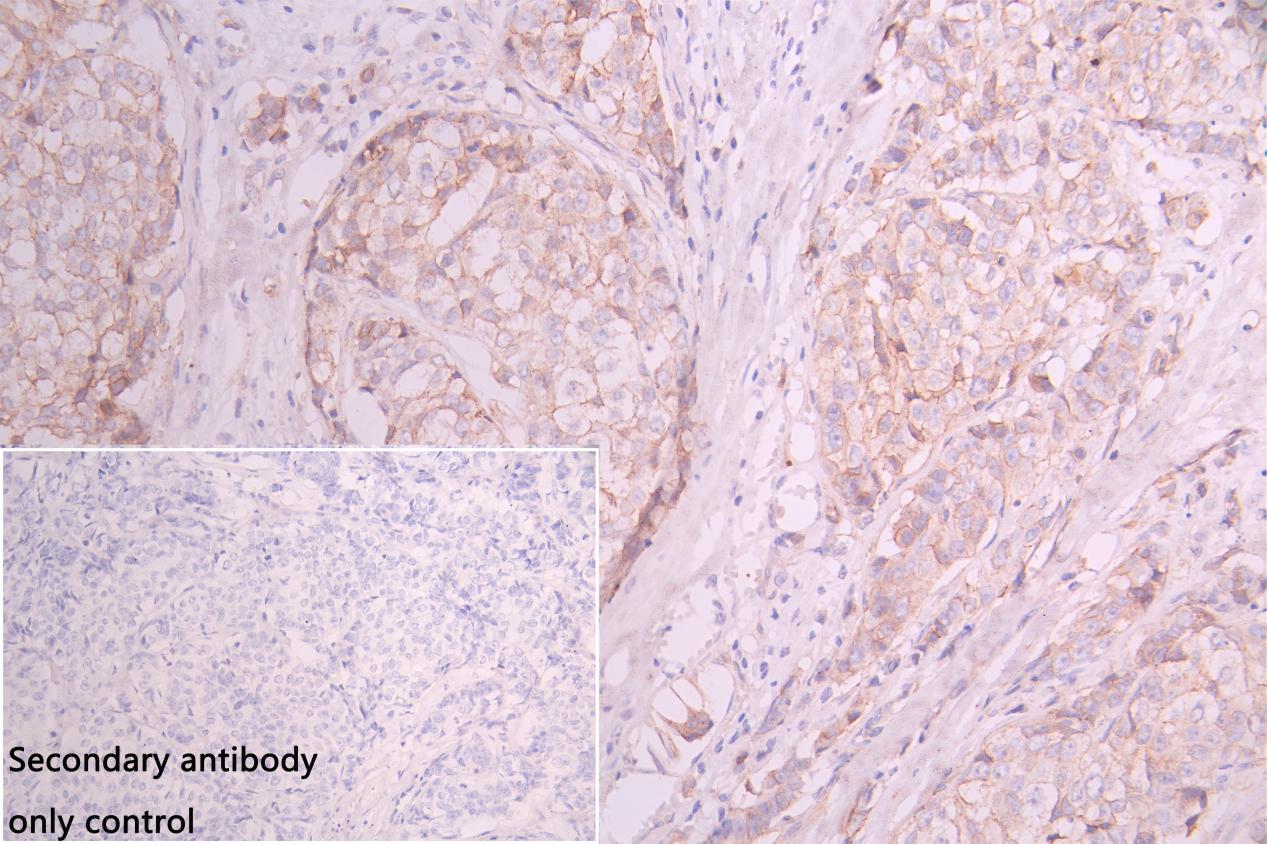
IHC image of CSB-PA015988LA01HU diluted at 1:100 and staining in paraffin-embedded human breast cancer performed on a Leica BondTM system. After dewaxing and hydration, antigen retrieval was mediated by high pressure in a citrate buffer (pH 6.0). Section was blocked with 10% normal goat serum 30min at RT. Then primary antibody (1% BSA) was incubated at 4C overnight. The primary is detected by a Goat anti-rabbit polymer IgG labeled by HRP and visualized using 0.05% DAB.Secondary antibody only control: uses 1% BSA instead of primary antibody |
|
|
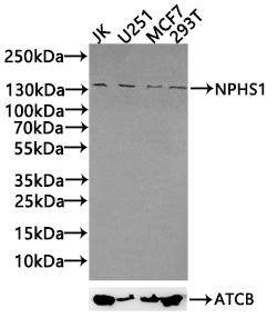
Positive WB detected in: JK whole cell lysate30µg), U251 whole cell lysate(30µg), MCF7 whole cell lysate(30µg), 293T whole cell lysate(30µg) All lanes: NPHS1 antibody at 1:1000 Secondary Goat polyclonal to rabbit IgG at 1/50000 dilution Predicted band size: 135,131 kDa Observed band size: 135 kDa Exposure time:120s |
Product Guarantee and Expert Support
