
Mono-methyl-H1F0 (K101) Antibody, Unconjugated, Rabbit, Polyclonal
Catalog Number:
CSB-PA010087OA101ME1HU
- Images (4)
| Article Name: | Mono-methyl-H1F0 (K101) Antibody, Unconjugated, Rabbit, Polyclonal |
| Biozol Catalog Number: | CSB-PA010087OA101ME1HU |
| Supplier Catalog Number: | CSB-PA010087OA101me1HU |
| Alternative Catalog Number: | CSB-PA010087OA101ME1HU-100UL, CSB-PA010087OA101ME1HU-50UL |
| Manufacturer: | Cusabio |
| Host: | Rabbit |
| Category: | Antikörper |
| Application: | ChIP, ELISA, ICC, IF, WB |
| Species Reactivity: | Human |
| Conjugation: | Unconjugated |
| Alternative Names: | H1 histone family member 0 antibody, H1(0) antibody, H10 antibody, H10_HUMAN antibody, h1f0 antibody, H1FV antibody, Histone H1 antibody, Histone H1(0) antibody, Histone H1.0 antibody, Histone H10 antibody, Histone H5 antibody, MGC5241 antibody, N-terminally processed antibody |
| Clonality: | Polyclonal |
| UniProt: | P07305 |
| Buffer: | Preservative: 0.03% Proclin 300<br />Constituents: 50% Glycerol, 0.01M PBS, pH 7.4 |
| Purity: | Antigen Affinity Purified |
| Form: | Liquid |
| Target: | H1F0 |
| Application Dilute: | Recommended dilution: WB:1:50-1:500, ICC:1:1-1:10, IF:1:1-1:10 |
|
|
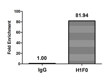
Chromatin Immunoprecipitation Hela (4*106) were treated with Micrococcal Nuclease, sonicated, and immunoprecipitated with 5µg anti-H1F0 (CSB-PA010087OA101me1HU) or a control normal rabbit IgG. The resulting ChIP DNA was quantified using real-time PCR with primers against the beta-Globin promoter. |
|
|
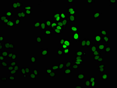
Immunofluorescence staining of HepG2 cells with CSB-PA010087OA101me1HU at 1:2.5, counter-stained with DAPI. The cells were fixed in 4% formaldehyde, permeabilized using 0.2% Triton X-100 and blocked in 10% normal Goat Serum. The cells were then incubated with the antibody overnight at 4°,C. The secondary antibody was Alexa Fluor 488-congugated AffiniPure Goat Anti-Rabbit IgG(H+L). |
|
|
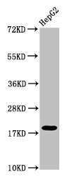
Western Blot Positive WB detected in: HepG2 whole cell lysate All lanes: H1F0 antibody at 1:50 Secondary Goat polyclonal to rabbit IgG at 1/50000 dilution Predicted band size: 21, 20 kDa Observed band size: 21 kDa |
|
|
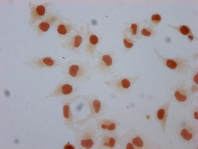
Immunocytochemistry analysis of CSB-PA010087OA101me1HU diluted at 1:5 and staining in Hela cells performed on a Leica BondTM system. The cells were fixed in 4% formaldehyde, permeabilized using 0.2% Triton X-100 and blocked with 10% normal goat serum 30min at RT. Then primary antibody (1% BSA) was incubated at 4°,C overnight. The primary is detected by a biotinylated secondary antibody and visualized using an HRP conjugated SP system. |
Product Guarantee and Expert Support
