0.01M TBS (pH7.4) with 1% rAlbumin, 0.02% Proclin300 and 50% Glycerol.
Form:
Liquid
Target:
PPP1R12A
Application Dilute:
WB=1:500-2000, Flow-Cyt=1µg /Test
Application Notes:
Modification: Phosphorylated
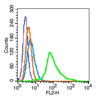
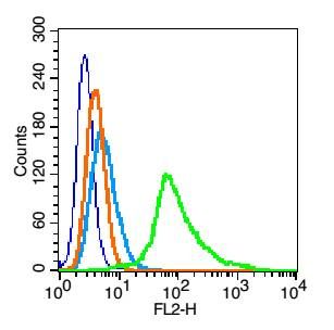
Blank control (blue): Hela cells (fixed with 2% paraformaldehyde (10 min), then permeabilized with 90% ice-cold methanol for 30 min on ice). Primary Antibody: Rabbit Anti-Phospho-MYPT1 (Thr696) antibody (orb6993), dilution: 1 µg in 100 µl 1X PBS containing 0.5% BSA, Isotype Control Antibody: Rabbit IgG (orange), used under the same conditions, Secondary Antibody: Goat anti-rabbit IgG-PE (white blue), dilution: 1:200 in 1X PBS containing 0.5% BSA.
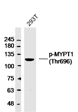
Sample: 293T (human) cell Lysate at 40 ug, Primary: Anti-p-MYPT1 (Thr696) (orb6993) at 1/300 dilution, Secondary: IRDye800CW Goat Anti-Rabbit IgG at 1/20000 dilution, Predicted band size: 113kD, Observed band size: 120 kD.
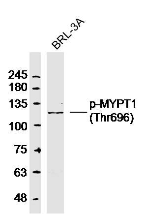
Sample: BRL-3A (Rat) cell Lysate at 40 ug, Primary: Anti-p-MYPT1 (Thr696) (orb6993) at 1/300 dilution, Secondary: IRDye800CW Goat Anti-Rabbit IgG at 1/20000 dilution, Predicted band size: 113kD, Observed band size: 120 kD.
Sample: Lane 1: Mouse Testis tissue lysates, Lane 2: Human HeLa cell lysates, Lane 3: Human MCF-7 cell lysates, Primary: Anti-Phospho-MYPT1 (Thr696) (orb6993) at 1/1000 dilution, Secondary: IRDye800CW Goat Anti-Rabbit IgG at 1/20000 dilution, Predicted band size: 113 kDa, Observed band size: 125 kDa.
Western blot analysis of BRL-3A (Rat)cell Lysate using MYPT1 (phospho-Thr696) antibody.
Western blot analysis of 293T (human)cell Lysate using MYPT1 (phospho-Thr696) antibody.
Flow cytometric analysis of Hela cells using MYPT1 (phospho-Thr696) antibody.
* VAT and and shipping costs not included. Errors and price changes excepted