E.coli-derived human KAP1 recombinant protein (Position: A699-P835). Human KAP1 shares 94.9% amino acid (aa) sequence identity with both mouse and rat KAP1.
Conjugation:
Unconjugated
Alternative Names:
KAP 1, KAP1, KRAB associated protein 1, KRAB interacting protein 1, KRIP 1, Nuclear corepressor KAP 1, RING finger protein 96, RNF96, TF1B, TIF1 beta, TIF1B, TRIM28, tripartite motif containing 28
Anti-KAP1/TRIM28 Antibody (monoclonal, 9E3). Tested in Flow Cytometry, IF, IHC, IHC-F, ICC, WB applications. This antibody reacts with Human, Mouse, Rat.
Western blot, 0.1-0.5µg/ml, Human, Mouse, Rat Immunohistochemistry (Paraffin-embedded Section), 0.5-1µg/ml, Human, Mouse, Rat Immunohistochemistry (Frozen Section), 0.5-1µg/ml, Human, Rat Immunocytochemistry/Immunofluorescence, 2µg/ml, Human Immunofluores
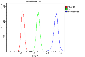
Flow Cytometry analysis of A549 cells using anti-KAP1/TRIM28 antibody. Overlay histogram showing A549 cells (Blue line). To facilitate intracellular staining, cells were fixed with 4% paraformaldehyde and permeabilized with permeabilization buffer. The cells were blocked with 10% normal goat serum. And then incubated with mouse anti-KAP1/TRIM28 Antibody (1 µg/1x10 6 cells) for 30 min at 20C. DyLight488 conjugated goat anti-mouse IgG (5-10 µg/1x10 6 cells) was used as secondary antibody for 30 minutes at 20C. Isotype control antibody (Green line) was mouse IgG (1 µg/1x10 6) used under the same conditions. Unlabelled sample without incubation with primary antibody and secondary antibody (Red line) was used as a blank control.
IF analysis of KAP1/TRIM28 using anti-KAP1/TRIM28 antibody. KAP1/TRIM28 was detected in immunocytochemical section of U20S cells. Enzyme antigen retrieval was performed using IHC enzyme antigen retrieval reagent for 15 mins. The cells were blocked with 10% goat serum. And then incubated with 2 µg/mL mouse anti-KAP1/TRIM28 Antibody overnight at 4C. DyLight488 Conjugated Goat Anti-Mouse IgG was used as secondary antibody at 1:100 dilution and incubated for 30 minutes at 37C. The section was counterstained with DAPI. Visualize using a fluorescence microscope and filter sets appropriate for the label used.
IF analysis of KAP1/TRIM28 using anti-KAP1/TRIM28 antibody. KAP1/TRIM28 was detected in paraffin-embedded section of human rectal cancer tissue. Heat mediated antigen retrieval was performed in EDTA buffer (pH8.0, epitope retrieval solution). The tissue section was blocked with 10% goat serum. The tissue section was then incubated with 2 µg/mL mouse anti-KAP1/TRIM28 Antibody overnight at 4C. DyLight488 Conjugated Goat Anti-Mouse IgG was used as secondary antibody at 1:100 dilution and incubated for 30 minutes at 37C. Visualize using a fluorescence microscope and filter sets appropriate for the label used.
IHC analysis of KAP1/TRIM28 using anti-KAP1/TRIM28 antibody. KAP1/TRIM28 was detected in frozen section of human placenta tissue. The tissue section was blocked with 10% goat serum. The tissue section was then incubated with 1 µg/ml mouse anti-KAP1/TRIM28 Antibody overnight at 4C. Biotinylated goat anti-mouse IgG was used as secondary antibody and incubated for 30 minutes at 37C. The tissue section was developed using Strepavidin-Biotin-Complex (SABC) with DAB as the chromogen.
IHC analysis of KAP1/TRIM28 using anti-KAP1/TRIM28 antibody. KAP1/TRIM28 was detected in paraffin-embedded section of human mammary cancer tissue. Heat mediated antigen retrieval was performed in EDTA buffer (pH8.0, epitope retrieval solution). The tissue section was blocked with 10% goat serum. The tissue section was then incubated with 1 µg/ml mouse anti-KAP1/TRIM28 Antibody overnight at 4C. Biotinylated goat anti-mouse IgG was used as secondary antibody and incubated for 30 minutes at 37C. The tissue section was developed using Strepavidin-Biotin-Complex (SABC) with DAB as the chromogen.
IHC analysis of KAP1/TRIM28 using anti-KAP1/TRIM28 antibody. KAP1/TRIM28 was detected in paraffin-embedded section of human rectal cancer tissue. Heat mediated antigen retrieval was performed in EDTA buffer (pH8.0, epitope retrieval solution). The tissue section was blocked with 10% goat serum. The tissue section was then incubated with 1 µg/ml mouse anti-KAP1/TRIM28 Antibody overnight at 4C. Biotinylated goat anti-mouse IgG was used as secondary antibody and incubated for 30 minutes at 37C. The tissue section was developed using Strepavidin-Biotin-Complex (SABC) with DAB as the chromogen.
IHC analysis of KAP1/TRIM28 using anti-KAP1/TRIM28 antibody. KAP1/TRIM28 was detected in paraffin-embedded section of rat intestine tissue. Heat mediated antigen retrieval was performed in EDTA buffer (pH8.0, epitope retrieval solution). The tissue section was blocked with 10% goat serum. The tissue section was then incubated with 1 µg/ml mouse an
* VAT and and shipping costs not included. Errors and price changes excepted