E.coli-derived human EIF6 recombinant protein (Position: N66-T210). Human EIF6 shares 99.3% amino acid (aa) sequence identity with both mouse and rat EIF6.
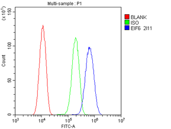
Flow Cytometry analysis of U20S cells using anti-EIF6 antibody. Overlay histogram showing U20S cells (Blue line). To facilitate intracellular staining, cells were fixed with 4% paraformaldehyde and permeabilized with permeabilization buffer. The cells were blocked with 10% normal goat serum. And then incubated with mouse anti-EIF6 Antibody (1 µg/1x10 6 cells) for 30 min at 20C. DyLight488 conjugated goat anti-mouse IgG (5-10 µg/1x10 6 cells) was used as secondary antibody for 30 minutes at 20C. Isotype control antibody (Green line) was mouse IgG (1 µg/1x10 6) used under the same conditions. Unlabelled sample (Red line) was also used as a control.
WB analysis using anti-EIF6 antibody.Lane 1:human placenta tissue, 2:
IHC analysis of EIF6 using anti-EIF6 antibody. EIF6 was detected in paraffin-embedded section of human intestinal cancer tissue. Heat mediated antigen retrieval was performed in EDTA buffer (pH8.0, epitope retrieval solution). The tissue section was blocked with 10% goat serum. The tissue section was then incubated with 1 µg/ml mouse anti-EIF6 Antibody overnight at 4C. Biotinylated goat anti-mouse IgG was used as secondary antibody and incubated for 30 minutes at 37C. The tissue section was developed using Strepavidin-Biotin-Complex (SABC) with DAB as the chromogen.
IHC analysis of EIF6 using anti-EIF6 antibody. EIF6 was detected in paraffin-embedded section of human lung cancer tissue. Heat mediated antigen retrieval was performed in EDTA buffer (pH8.0, epitope retrieval solution). The tissue section was blocked with 10% goat serum. The tissue section was then incubated with 1 µg/ml mouse anti-EIF6 Antibody overnight at 4C. Biotinylated goat anti-mouse IgG was used as secondary antibody and incubated for 30 minutes at 37C. The tissue section was developed using Strepavidin-Biotin-Complex (SABC) with DAB as the chromogen.
IHC analysis of EIF6 using anti-EIF6 antibody. EIF6 was detected in paraffin-embedded section of human mammary cancer tissue. Heat mediated antigen retrieval was performed in EDTA buffer (pH8.0, epitope retrieval solution). The tissue section was blocked with 10% goat serum. The tissue section was then incubated with 1 µg/ml mouse anti-EIF6 Antibody overnight at 4C. Biotinylated goat anti-mouse IgG was used as secondary antibody and incubated for 30 minutes at 37C. The tissue section was developed using Strepavidin-Biotin-Complex (SABC) with DAB as the chromogen.
IHC analysis of EIF6 using anti-EIF6 antibody. EIF6 was detected in paraffin-embedded section of rat spleen tissue. Heat mediated antigen retrieval was performed in EDTA buffer (pH8.0, epitope retrieval solution). The tissue section was blocked with 10% goat serum. The tissue section was then incubated with 1 µg/ml mouse anti-EIF6 Antibody overnight at 4C. Biotinylated goat anti-mouse IgG was used as secondary antibody and incubated for 30 minutes at 37C. The tissue section was developed using Strepavidin-Biotin-Complex (SABC) with DAB as the chromogen.
Western blot analysis of EIF6 using anti-EIF6 antibody. Electrophoresis was performed on a 5-20% SDS-PAGE gel at 70V (Stacking gel) / 90V (Resolving gel) for 2-3 hours. The sample well of each lane was loaded with 50 ug of sample under reducing conditions. Lane 1: human placenta tissue lysates, Lane 2: human CACO-2 whole cell lysates, Lane 3: human HEPG2 whole cell lysates, Lane 4: human K562 whole cell lysates, Lane 5: human A549 whole cell lysates, Lane 6: human PC-3 whole cell lysates, Lane 7: human Hela whole cell lysates, Lane 8: human U87 whole cell lysates, Lane 9: rat kidney tissue lysates, Lane 10: rat testis tissue lysates, Lane 11: mouse HEPA1-6 whole cell lysates. After Electrophoresis, proteins were transferred to a Nitrocellulose membrane at 150mA for 50-90 minutes. Blocked the membrane with 5% Non-fat Milk/ TBS for 1.5 hour at RT. The membrane was incubated with mouse anti-EIF6 antigen affinity purified monoclonal antibody at 0.5 µg/mL overnight at 4C, then washed with TBS-0.1% Tween 3 times with 5 minutes each and probed with a go
* VAT and and shipping costs not included. Errors and price changes excepted