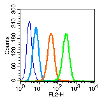
Flow cytometric analysis of MCF7 tissue using FSH receptor antibody
Immunohistochemical staining of paraffin embedded rat ovary tissue using FSH receptor antibody
Blank control (blue line): MCF7 (fixed with 70% methanol overnight at 4C). Primary Antibody (green line): Rabbit Anti-FSH receptor antibody (orb500733), dilution: 0.2 µg/10 6 cells, Isotype Control Antibody (orange line): Rabbit IgG. Secondary Antibody (white blue line): Goat anti-rabbit IgG-PE, dilution: 1 µg/Test.
Paraformaldehyde-fixed, paraffin embedded (rat ovary), Antigen retrieval by boiling in sodium citrate buffer (pH6.0) for 15 min, Block endogenous peroxidase by 3% hydrogen peroxide for 20 minutes, Blocking buffer (normal goat serum) at 37C for 30 min, Antibody incubation with (FSH receptor) Polyclonal Antibody, Unconjugated (orb500733) at 1:400 overnight at 4C, followed by a conjugated secondary antibody for 20 minutes and DAB staining.
Paraformaldehyde-fixed, paraffin embedded (rat testis), Antigen retrieval by boiling in sodium citrate buffer (pH6.0) for 15 min, Block endogenous peroxidase by 3% hydrogen peroxide for 20 minutes, Blocking buffer (normal goat serum) at 37C for 30 min, Antibody incubation with (CBFb) Polyclonal Antibody, Unconjugated (orb500733) at 1:200 overnight at 4C, followed by operating according to SP Kit (Rabbit) instructionsand DAB staining.
Sample: Ovary (mouse) Lysate at 40 ug, Primary: Anti-FSH receptor (orb500733) at 1/300 dilution, Secondary: IRDye800CW Goat Anti-Rabbit IgG at 1/20000 dilution, Predicted band size: 78kD, Observed band size: 78kD.
* VAT and and shipping costs not included. Errors and price changes excepted