Phosphate buffered saline (PBS), pH 7.4, 15 mM sodium azide
Target:
gamma-Tubulin
Application Notes:
Application Notes: Immunocytochemistry: Recommended dilution: 1-2 µg/ml. Staining technique: (a) Fix cells for 10 min in methanol at -20C and for 6 min in acetone at -20C, (b) Fix cells directly in methanol for 10 min at -20C or in acetone for 10 min at -20C. Positive control: P-19 murine embryonal carcinoma cell line, 3T3 murine fibroblasts. The antibody TU-30 stains only fixed cells.Western blotting: Recommended dilution 1-2 µg/ml, reducing conditions
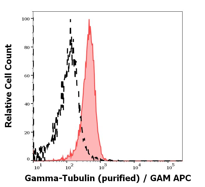
Separation of MCF-7 cells stained using anti-gamma-Tubulin (TU-30) purified antibody (concentration in sample 9 µg/ml, GAM APC, red-filled) from MCF-7 cells unstained by primary antibody (GAM APC, black-dashed) in flow cytometry analysis (intracellular staining).
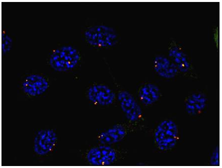
Immunocytochemistry staining of P19X1 mouse embryonal carcinoma cell line using anti-gamma-tubulin (TU-30) (detection by secondary antibody Goat anti-mouse Cy3). Nuclei were stained with DAPI (blue).
Immunocytochemistry staining of murine fibroblasts using anti-gamma-tubulin (TU-30, direct conjugate with Dyomics 547, red). Nuclei were stained with DAPI (blue).
Immunocytochemistry staining of microtubular networks in 3T3 mouse fibroblasts. A - metaphase, B - anaphase, C - telophase. Gamma-tubulin (red) stained with anti-gamma-tubulin (TU-30), alpha-tubulin (green) with polyclonal anti-alpha-tubulin antibody and nuclei with DAPI (blue).
Western blotting analysis of human gamma-tubulin using mouse monoclonal antibody TU-30 on lysates of various cell lines under reducing and non-reducing conditions. Nitrocellulose membrane was probed with 2 µg/ml of mouse anti-gamma-tubulin monoclonal antibody followed by IRDye800-conjugated anti-mouse secondary antibody. A specific band was detected for gamma-tubulin at approximately 46 kDa.
Western blotting analysis of differential reactivity of monoclonal antibodies to gamma-tubulin with human gamma-tubulin isotypes. (A) Immunoblots of total cell lysates from SH-SY5Y cells, expressing TagRFP-tagged human gamma-tubulin 1 (gamma-Tb1) or gamma-tubulin 2 (gamma-Tb2), probed with Abs to gamma-tubulin (TU-30, TU-32), TagRFP (RFP) and GAPDH. In control samples, only secondary anti-mouse Ab was applied. (B) Immunoblots of immobilized GST-tagged human C-terminal regions (a.a. 362-451) of gamma-Tb1 or gamma-Tb2 probed with Abs to gamma-tubulin (TU-30, TU-32) and GST. In control samples, only secondary anti-mouse Ab was applied.
* VAT and and shipping costs not included. Errors and price changes excepted