Phosphate buffered saline (PBS), pH 7.4, 15 mM sodium azide
Target:
betaIII-Tubulin
Application Notes:
Application Notes: Western blotting: Recommended dilution: 1-2 µg/ml, positive control: porcine brain lysate, negative control: HPB-ALL human peripheral blood leukemia cell line, reducing conditions. Immunohistochemistry (paraffin sections): Recommended dilution: 10 µg/ml, standard ABC technique (DAB+), pretreatment: 0.1% pepsin (trypsin) in 0.1 M HCl, incubation 30 min in RT, or high temperature citrate buffer antigen retrieval, positive tissue: neuronal tissue. Immunocytochemistry: Positive material: Neuro2a mouse neuroblastoma cell line. Flow cytometry: Recommended dilution: 1-4 µg/ml. Intracellular staining
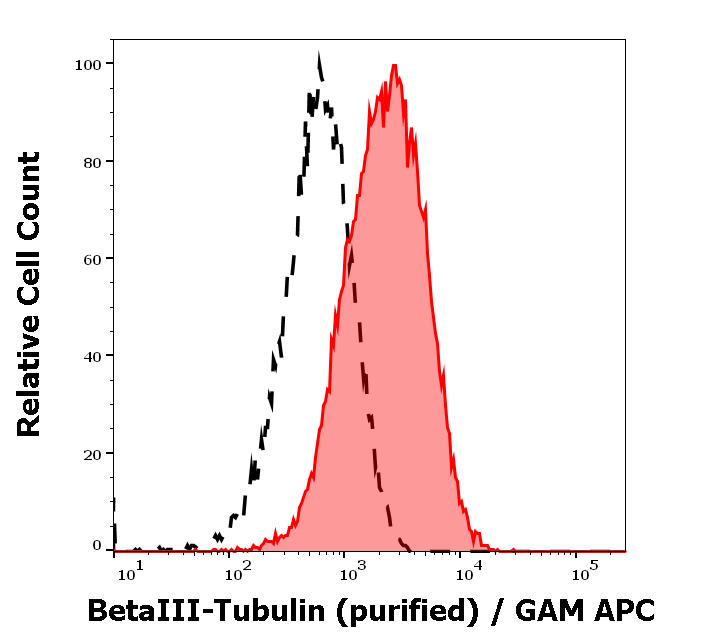
Separation of MCF-7 cells stained using anti-betaIII-Tubulin (TU-20) purified antibody (concentration in sample 9 µg/ml, GAM APC, red-filled) from MCF-7 cells unstained by primary antibody (GAM APC, black-dashed) in flow cytometry analysis (intracellular staining).
Immunocytochemistry staining of Neuro2a mouse neuroblastoma cell line using anti-betaIII-tubulin (TU-20, green, 3 µg/ml). Nuclei were stained with DAPI (blue).
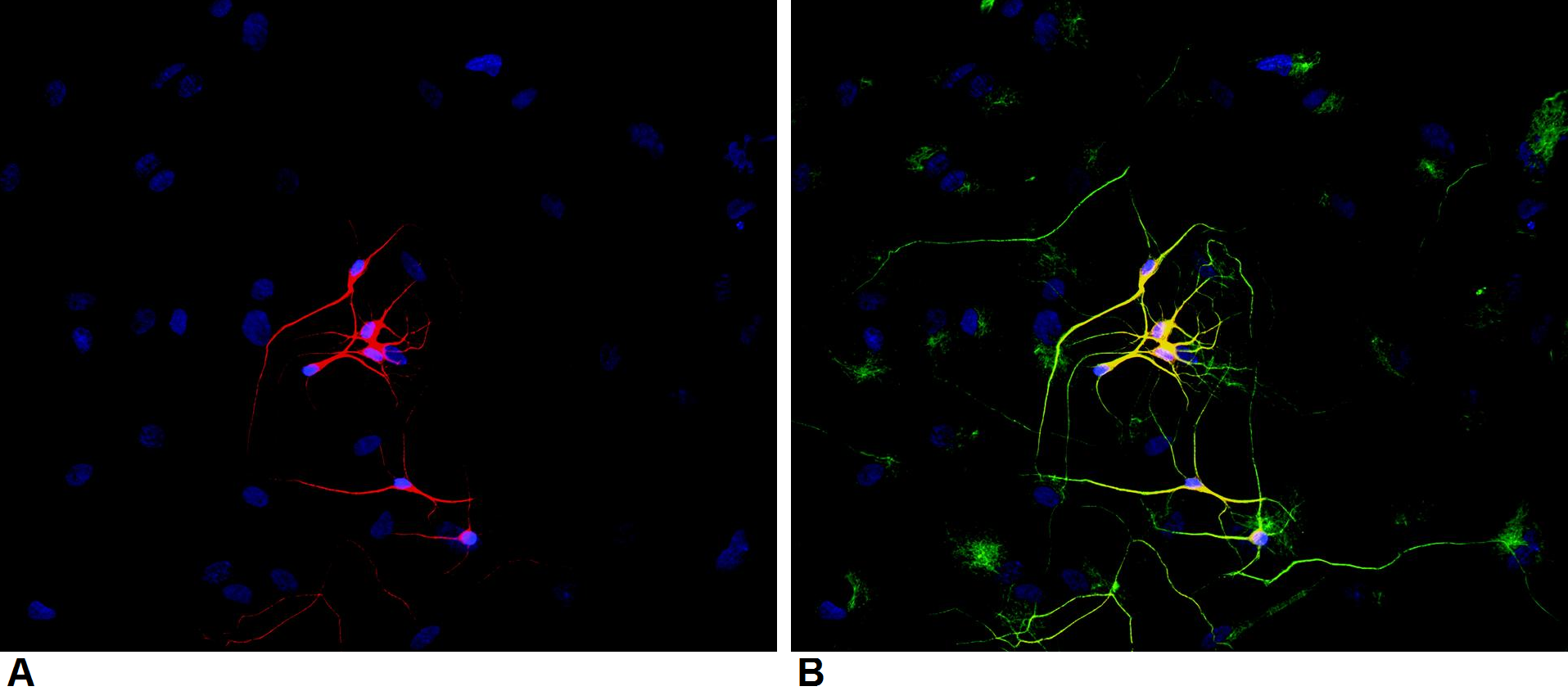
Immunocytochemistry staining of P-19 mouse embryonal carcinoma cell line stimulated to neuronal differentiation by retinoic acid. A - Microtubules decorated with neuron-specific anti-betaIII-tubulin (TU-20, red). B - Merged image of co-staining with anti-beta-tubulin (TU-06, green, cat. no. orb44535). Superposition of red and green colours provided yellow staining. Nuclei were stained with DNA-binding dye (blue).
Immunohistochemistry staining of human brain (paraffin sections) using anti-betaIII tubulin (TU-20).
Immunohistochemistry staining of betaIII tubulin (red) in tissue sections of murine brain expressing GFP in some of its neurons (green). Mouse monoclonal antibody TU-20 (anti-betaIII tubulin) was detected with goat anti-mouse IgG1 conjugated with Alexa Fluor 555.
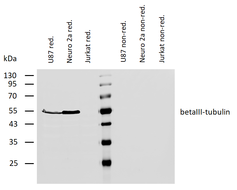
Western blotting analysis of human betaIII-tubulin using mouse monoclonal antibody TU-20 on lysates of U87 cells and Neuro 2a cells (and Jurkat cells as a negative control) under reducing and non-reducing conditions. Nitrocellulose membrane was probed with 2 µg/ml of mouse anti-betaIII-tubulin monoclonal antibody followed by IRDye800-conjugated anti-mouse secondary antibody. A specific band was detected for betaIII-tubulin at approximately 55 kDa.
* VAT and and shipping costs not included. Errors and price changes excepted