Tris buffered saline (TBS), pH 8.0, 15 mM sodium azide
Target:
alpha-Tubulin
Application Notes:
Application Notes: Immunohistochemistry (paraffin sections): Recommended dilution: 10 µg/ml. Immunoprecipitation: Reducing conditions.Western blotting: Recommended dilution: 1-2 µg/ml. This antibody can be used for Western blotting, but its alternative TU-02 (orb44534) gives better signal in this application
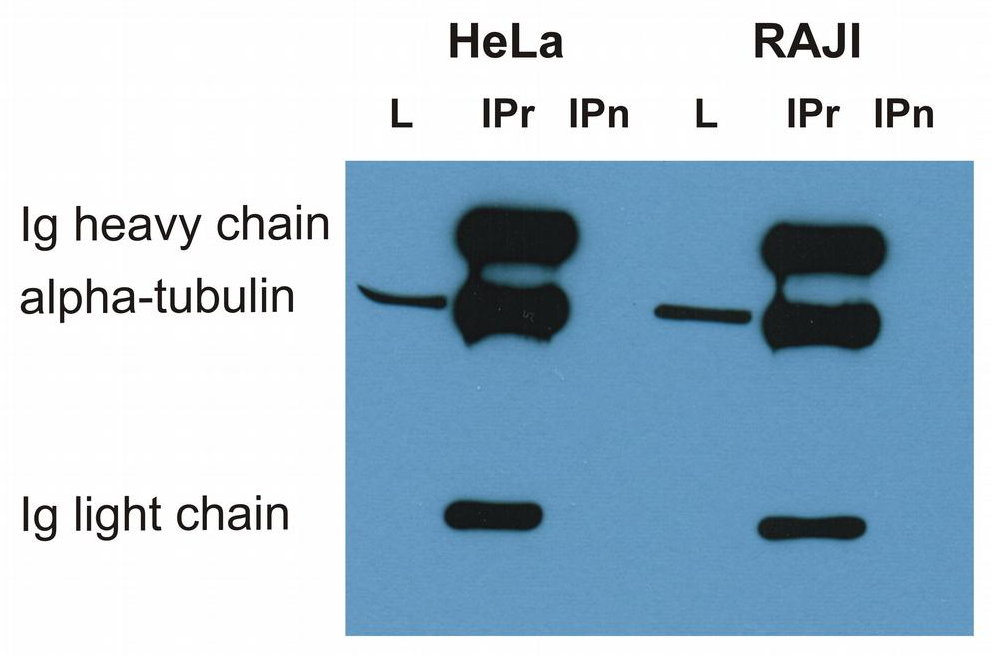
Immunoprecipitation of alpha-tubulin from HeLa and RAJI cell lysate by antibody TU-16 and its detection by antibody TU-01. IgM heavy chain (76-92 kDa) and IgM light chain (25-30 kDa) indicated. Mr of alpha tubulin is around 50 kDa. L = lysate, IPr = immunoprecipitate (reducing conditions).
Immunocytochemistry staining of alpha-tubulin in Hep-2 cells using mouse monoclonal antibody TU-16 (diluted 1:400), detected with GAM IgG-Alexa Fluor488 (diluted 1:200, green).
Immunohistochemistry staining of human heart (paraffin sections) using anti-alpha tubulin (TU-16).
Immunohistochemistry staining (paraffin sections) of alpha-tubulin in human stomach using mouse monoclonal antibody TU-16 (diluted 1:400), detected with GAM IgG-Alexa Fluor488 (diluted 1:200, green).
Western blotting analysis of human alpha-tubulin using mouse monoclonal antibody TU-16 on lysates of various cell lines and porcine brain under reducing and non-reducing conditions. Nitrocellulose membrane was probed with 2 µg/ml of mouse anti-alpha-tubulin monoclonal antibody followed by IRDye800-conjugated anti-mouse secondary antibody. A specific band was detected for alpha-tubulin at approximately 54 kDa, nonspecific minor bands above 100 kDa do not interfere with specific signal.
Anti-alpha-Tubulin Purified (TU-16) works in WB application under reducing conditions. Western blotting analysis was performed on whole cell extracts (RIPA lysis buffer) of HeLa, HEK 293, ESS-1 and Jurkat cell lines mixed and heated (100C, 5 min) with reducing (2-mercaptoethanol) or non-reducing SDS-loading buffer. Samples were resolved using 12% Tris-glycine SDS gel electrophoresis. Nitrocellulose membrane blot was probed simultaneously with mouse IgM monoclonal antibody TU-16 (1 µg/ml) and mouse IgG1 anti-GAPDH monoclonal antibody FF26A (1 µg/ml) used as the loading control. Subclass-specific secondary antibodies IRDye 680RD Goat-anti-Mouse IgM (red) and IRDye 800CW Goat-anti-Mouse IgG (green) were used for multiplex fluorescent Western blot detection. Alpha-tubulin was detected at ~50 kDa in all tested cell lines.
* VAT and and shipping costs not included. Errors and price changes excepted