This TPK1 Antibody is an unconjugated polyclonal product. It tagets TPK1 using a Recombinant Human Thiamin pyrophosphokinase 1 protein (1-243AA) as the immunogen. This antibody is suitable for ELISA, IF, IHC, IP, WB. Purification: Antigen Affinity Purified.
Positive WB detected in: U251 whole cell lysate(30µg), THP-1 whole cell lysate(30µg), MCF-7 whole cell lysate(30µg), K562 whole cell lysate(30µg), CT-26 whole cell lysate(30µg), PC-3 whole cell lysate(30µg), JK whole cell lysate(30µg), Mouse Liver tissue lysate(30µg), Mouse Kidneyr tissue lysate(30µg). All lanes: TPK1 antibody at 1:1000. Secondary: Goat polyclonal to rabbit IgG at 1/40000 dilution. Predicted band size: 27,14kDa, Observed band size: 27 kDa, Exposure time: 180s.
IHC image of orb41306 diluted at 1:100 and staining in paraffin-embedded human Kidney tissue performed on a Leica BondTM system. After dewaxing and hydration, antigen retrieval was mediated by high pressure in a citrate buffer (pH 6.0). Section was blocked with 10% normal goat serum 30min at RT. Then primary antibody (1% BSA) was incubated at 4C overnight. The primary is detected by a Goat anti-rabbit polymer IgG labeled by HRP and visualized using 0.05% DAB. Secondary antibody only control: uses 1% BSA instead of primary antibody.
IHC image of orb41306 diluted at 1:100 and staining in paraffin-embedded human Heart tissue performed on a Leica BondTM system. After dewaxing and hydration, antigen retrieval was mediated by high pressure in a citrate buffer (pH 6.0). Section was blocked with 10% normal goat serum 30min at RT. Then primary antibody (1% BSA) was incubated at 4C overnight. The primary is detected by a Goat anti-rabbit polymer IgG labeled by HRP and visualized using 0.05% DAB. Secondary antibody only control: uses 1% BSA instead of primary antibody.
IHC image of orb41306 diluted at 1:100 and staining in paraffin-embedded human breast cancer performed on a Leica BondTM system. After dewaxing and hydration, antigen retrieval was mediated by high pressure in a citrate buffer (pH 6.0). Section was blocked with 10% normal goat serum 30min at RT. Then primary antibody (1% BSA) was incubated at 4C overnight. The primary is detected by a Goat anti-rabbit polymer IgG labeled by HRP and visualized using 0.05% DAB. Secondary antibody only control: uses 1% BSA instead of primary antibody.
Immunofluorescence analysis of PC3 cells using TPK1 antibody
Immunohistochemical staining of human kidney tissue using TPK1 antibody
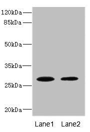
Western blot analysis of Mouse liver(lane 1), Mouse kidney(lane 2) tissue using TPK1 antibody
* VAT and and shipping costs not included. Errors and price changes excepted