Purified polyclonal antibody supplied in PBS with 0.09% (W/V) sodium azide. This antibody is purified through a protein A column, followed by peptide affinity purification.
Immunofluorescense analysis of 293 cell using ESRRA antibody (primary antibody dilution at: 1:10-50)
Western blot analysis of ESRRA Antibody (Center) in A375 cell line lysates (35 ug/lane). ESRRA (arrow) was detected using the purified Pab.
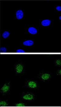
Confocal immunofluorescent analysis of ESRRA Antibody (Center) with 293 cell followed by Alexa Fluor 488-conjugated goat anti-rabbit lgG (green). DAPI was used to stain the cell nuclear (blue).
Western blot analysis of ESRRA (arrow) using rabbit polyclonal ESRRA Antibody (Center). 293 cell lysates (2 ug/lane) either nontransfected (Lane 1) or transiently transfected (Lane 2) with the ESRRA gene.
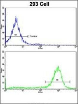
Flow cytometric analysis of 293 cells using ESRRA Antibody (Center) (bottom histogram) compared to a negative control cell (top histogram). FITC-conjugated goat-anti-rabbit secondary antibodies were used for the analysis.
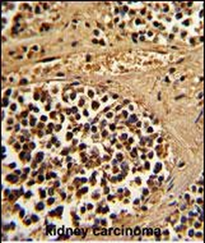
Formalin-fixed and paraffin-embedded human kidney carcinoma reacted with ESRRA Antibody (Center), which was peroxidase-conjugated to the secondary antibody, followed by DAB staining. This data demonstrates the use of this antibody for immunohistochemistry, clinical relevance has not been evaluated.
* VAT and and shipping costs not included. Errors and price changes excepted