Purified polyclonal antibody supplied in PBS with 0.09% (W/V) sodium azide. This antibody is prepared by Saturated Ammonium Sulfate (SAS) precipitation followed by dialysis against PBS.
Target:
NEUROD1
Application Dilute:
WB: 1:1000, IF/ICC: 1:100, IHC-P: 1:50-100
Immunofluorescense analysis of ES cells using NeuroD1 antibody (primary antibody dilution at: 1:100)
orb33616
Western blot analysis of hNeuroD1-Q30 in HepG2 cell line lysates (35 ug/lane). NEUROD1 (arrow) was detected using the purified Pab.
Formalin-fixed and paraffin-embedded human cancer tissue reacted with the primary antibody, which was peroxidase-conjugated to the secondary antibody, followed by DAB staining. This data demonstrates the use of this antibody for immunohistochemistry, clinical relevance has not been evaluated. BC = breast carcinoma, HC = hepatocarcinoma.
ES cells were transiently transfected with flag-tagged mouse NeuroD1 (tagged on N-term). Fixed 24h post transfection.Stained for flag tag (red) to check that some cells express protein. Most protein was in nucleus but some was cytoplasmic. Stained with NeuroD1 N-term antibodies at 1:100. NeuroD1 N-term antibody showed strong and clear staining with similar pattern to the flag staining.
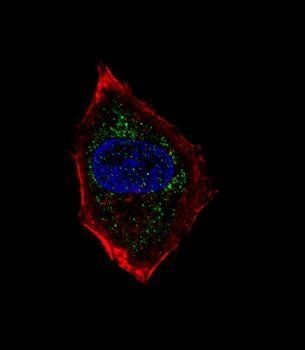
Fluorescent confocal image of HepG2 cell stained with hNeuroD1-Q30. HepG2 cells were fixed with 4% PFA (20 min), permeabilized with Triton X-100 (0.1%, 10 min), then incubated with hNeuroD1-Q30 primary antibody (1:25, 1 h at 37C). For secondary antibody, Alexa Fluor 488 conjugated donkey anti-rabbit antibody (green) was used (1:400, 50 min at 37C). Cytoplasmic actin was counterstained with Alexa Fluor 555 (red) conjugated Phalloidin (7units/ml, 1 h at 37C). Nuclei were counterstained with DAPI (blue) (10 µg/ml, 10 min). hNeuroD1-Q30 immunoreactivity is localized to vesicles significantly.
* VAT and and shipping costs not included. Errors and price changes excepted