A synthetic peptide corresponding to a sequence at the C-terminus of human PDPK1, different from the related mouse and rat sequences by two amino acids.
Conjugation:
Unconjugated
Alternative Names:
3-phosphoinositide-dependent protein kinase 1, hPDK1, 2.7.11.1, PDPK1, PDK1
PDPK1 Rabbit Polyclonal Antibody
Clonality:
Polyclonal
Concentration:
Adding 0.2 ml of distilled water will yield a concentration of 500 µg/ml.
Each vial contains antibody formulated with stabilizing components, 0.9 mg NaCl, 0.2 mg Na2HPO4, and 0.05 mg NaN3. *This antibody is supplied in a stabilized formulation. Compatibility with conjugation reactions depends on the chemistry of the conjugation
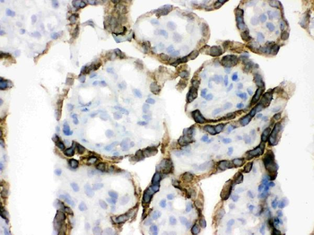
IHC analysis of PDPK1 using anti-PDPK1 antibody. PDPK1 was detected in frozen section of human placenta tissue. The tissue section was blocked with 10% goat serum. The tissue section was then incubated with 1 µg/ml rabbit anti-PDPK1 Antibody overnight at 4C. Biotinylated goat anti-rabbit IgG was used as secondary antibody and incubated for 30 minutes at 37C. The tissue section was developed using Strepavidin-Biotin-Complex (SABC) with DAB as the chromogen.
IHC analysis of PDPK1 using anti-PDPK1 antibody.PDPK1 was detected in paraffin-embedded section
IHC analysis of PDPK1 using anti-PDPK1 antibody. PDPK1 was detected in frozen section of mouse small intestine tissue. The tissue section was blocked with 10% goat serum. The tissue section was then incubated with 1 µg/ml rabbit anti-PDPK1 Antibody overnight at 4C. Biotinylated goat anti-rabbit IgG was used as secondary antibody and incubated for 30 minutes at 37C. The tissue section was developed using Strepavidin-Biotin-Complex (SABC) with DAB as the chromogen.
IHC analysis of PDPK1 using anti-PDPK1 antibody. PDPK1 was detected in paraffin-embedded section of Human Lung Cancer Tissue. Heat mediated antigen retrieval was performed in citrate buffer (pH6, epitope retrieval solution) for 20 mins. The tissue section was blocked with 10% goat serum. The tissue section was then incubated with 1 µg/ml rabbit anti-PDPK1 Antibody overnight at 4C. Biotinylated goat anti-rabbit IgG was used as secondary antibody and incubated for 30 minutes at 37C. The tissue section was developed using Strepavidin-Biotin-Complex (SABC) with DAB as the chromogen.
IHC analysis of PDPK1 using anti-PDPK1 antibody. PDPK1 was detected in paraffin-embedded section of Mouse Intestine Tissue. Heat mediated antigen retrieval was performed in citrate buffer (pH6, epitope retrieval solution) for 20 mins. The tissue section was blocked with 10% goat serum. The tissue section was then incubated with 1 µg/ml rabbit anti-PDPK1 Antibody overnight at 4C. Biotinylated goat anti-rabbit IgG was used as secondary antibody and incubated for 30 minutes at 37C. The tissue section was developed using Strepavidin-Biotin-Complex (SABC) with DAB as the chromogen.
IHC analysis of PDPK1 using anti-PDPK1 antibody. PDPK1 was detected in paraffin-embedded section of Rat Testis Tissue. Heat mediated antigen retrieval was performed in citrate buffer (pH6, epitope retrieval solution) for 20 mins. The tissue section was blocked with 10% goat serum. The tissue section was then incubated with 1 µg/ml rabbit anti-PDPK1 Antibody overnight at 4C. Biotinylated goat anti-rabbit IgG was used as secondary antibody and incubated for 30 minutes at 37C. The tissue section was developed using Strepavidin-Biotin-Complex (SABC) with DAB as the chromogen.
Western blot analysis of PDPK1 using anti-PDPK1 antibody. Electrophoresis was performed on a 5-20% SDS-PAGE gel at 70V (Stacking gel) / 90V (Resolving gel) for 2-3 hours. The sample well of each lane was loaded with 50 ug of sample under reducing conditions. Lane 1: Rat Liver Tissue Lysate, Lane 2: Rat Lung Tissue Lysate, Lane 3: Mouse Liver Tissue Lysate, Lane 4: Mouse Lung Tissue Lysate, Lane 5: COLO320 Whole Cell Lysate, Lane 6: MCF-7 Whole Cell Lysate. After Electrophoresis, proteins were transferred to a Nitrocellulose membrane at 150mA for 50-90 minutes. Blocked the membrane with 5% Non-fat Milk/ TBS for 1.5 hour at RT. The membrane was incubated with rabbit anti-PDPK1 antigen affinity purified polyclonal antibody at 0.5 µg/mL overnight at 4C, then washed with TBS-0.1% Tween 3 times with 5 minutes each and probed with a goat anti-rabbit IgG-HRP secondary antibody at a dilution of 1:10000 for 1.5 hour at RT. The signal is developed using an Enhanced Chemiluminescent detection (ECL) kit with Tanon 5200 system. A specific band was detected for PDPK1 at approximately 70 KD. The expected band size for PDPK1 is at 63 KD.
* VAT and and shipping costs not included. Errors and price changes excepted