E.coli-derived human SDHA recombinant protein (Position: S44-L380). Human SDHA shares 98.2% and 97.6% amino acid (aa) sequence identity with mouse and rat SDHA, respectively.
Conjugation:
Unconjugated
Alternative Names:
Succinate dehydrogenase [ubiquinone] flavoprotein subunit, mitochondrial, 1.3.5.1, Flavoprotein subunit of complex II, Fp, SDHA, SDH2, SDHF
Anti-SDHA Antibody. Tested in Flow Cytometry, IF, IHC, ICC, WB applications. This antibody reacts with Human, Mouse, Rat.
Clonality:
Polyclonal
Concentration:
Adding 0.2 ml of distilled water will yield a concentration of 500 µg/ml.
Western blot, 0.1-0.5 µg/ml, Human, Mouse, Rat Immunohistochemistry (Paraffin-embedded Section), 2-5 µg/ml, Human, Mouse Immunocytochemistry/Immunofluorescence, 5 µg/ml, Human Immunoprecipitation, 0.5-2 µg/ml, Human Flow Cytometry(Fixed), 1-3 µg/1x10 6 ce
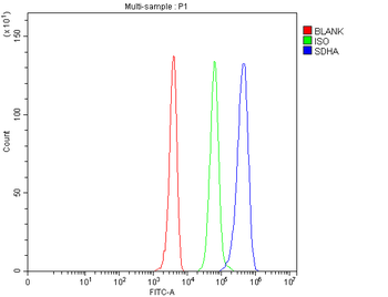
Flow Cytometry analysis of 293T cells using anti-SDHA antibody. Overlay histogram showing 293T cells (Blue line). To facilitate intracellular staining, cells were fixed with 4% paraformaldehyde and permeabilized with permeabilization buffer. The cells were blocked with 10% normal goat serum. And then incubated with rabbit anti-SDHA Antibody (1 µg/1x10 6 cells) for 30 min at 20C. DyLight488 conjugated goat anti-rabbit IgG (5-10 µg/1x10 6 cells) was used as secondary antibody for 30 minutes at 20C. Isotype control antibody (Green line) was rabbit IgG (1 µg/1x10 6) used under the same conditions. Unlabelled sample without incubation with primary antibody and secondary antibody (Red line) was used as a blank control.
IF analysis of SDHA using anti-SDHA antibody. SDHA was detected in an immunocytochemical section of U2OS cells. Enzyme antigen retrieval was performed using IHC enzyme antigen retrieval reagent for 15 mins. The cells were blocked with 10% goat serum. And then incubated with 5 µg/mL rabbit anti-SDHA Antibody overnight at 4C. DyLight488 Conjugated Goat Anti-Rabbit IgG was used as secondary antibody at 1:500 dilution and incubated for 30 minutes at 37C. The section was counterstained with DAPI. Visualize using a fluorescence microscope and filter sets appropriate for the label used.
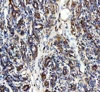
IHC analysis of SDHA using anti-SDHA antibody. SDHA was detected in a paraffin-embedded section of human breast cancer tissue. Heat mediated antigen retrieval was performed in EDTA buffer (pH8.0, epitope retrieval solution). The tissue section was blocked with 10% goat serum. The tissue section was then incubated with 2 µg/ml rabbit anti-SDHA Antibody overnight at 4C. Peroxidase Conjugated Goat Anti-rabbit IgG was used as secondary antibody and incubated for 30 minutes at 37C. The tissue section was developed using HRP Conjugated Rabbit IgG Super Vision Assay Kit with DAB as the chromogen.
IHC analysis of SDHA using anti-SDHA antibody. SDHA was detected in a paraffin-embedded section of human ovarian cancer tissue. Heat mediated antigen retrieval was performed in EDTA buffer (pH8.0, epitope retrieval solution). The tissue section was blocked with 10% goat serum. The tissue section was then incubated with 2 µg/ml rabbit anti-SDHA Antibody overnight at 4C. Peroxidase Conjugated Goat Anti-rabbit IgG was used as secondary antibody and incubated for 30 minutes at 37C. The tissue section was developed using HRP Conjugated Rabbit IgG Super Vision Assay Kit with DAB as the chromogen.
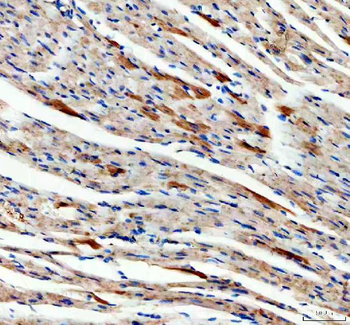
IHC analysis of SDHA using anti-SDHA antibody. SDHA was detected in a paraffin-embedded section of mouse heart tissue. Heat mediated antigen retrieval was performed in EDTA buffer (pH8.0, epitope retrieval solution). The tissue section was blocked with 10% goat serum. The tissue section was then incubated with 2 µg/ml rabbit anti-SDHA Antibody overnight at 4C. Peroxidase Conjugated Goat Anti-rabbit IgG was used as secondary antibody and incubated for 30 minutes at 37C. The tissue section was developed using HRP Conjugated Rabbit IgG Super Vision Assay Kit with DAB as the chromogen.
Western blot analysis of SDHA using anti-SDHA antibody. Electrophoresis was performed on a 5-20% SDS-PAGE gel at 70V (Stacking gel) / 90V (Resolving gel) for 2-3 hours. The sample well of each lane was loaded with 30 ug of sample under reducing conditions. Lane 1: human 293T whole cell lysates, Lane 2: human MCF-7 whole cell lysates, Lane 3: human HepG2 whole cell lysates, Lane 4: human Hela whole cell lysates, Lane 5: rat brain tissue lysates, Lane 6: rat kidney tissue lysates, Lane 7: mouse brain tissue lysates, Lane 8: mouse kidney tissue lysates. After electrophoresis, proteins were transferred to a nitrocellulose membrane at 150 mA for 50-90 minutes. Blocked the membrane with 5% non-fat milk/TBS for 1.5 hour at RT. The membrane was incubated with rabbit anti-SDHA antigen affinity purified polyclonal antibody at 0.5 µg/mL overnight at 4C, then washed with TBS-0.1% Tween 3 tim
* VAT and and shipping costs not included. Errors and price changes excepted