Each vial contains antibody formulated with stabilizing components, 0.9 mg NaCl, 0.2 mg Na2HPO4, and 0.05 mg NaN3. *This antibody is supplied in a stabilized formulation. Compatibility with conjugation reactions depends on the chemistry of the conjugation
Form:
Lyophilized
Target:
Pro-interleukin-16 [Cleaved into: Interleukin-16
Application Dilute:
Immunohistochemistry (Paraffin-embedded Section), 0.5-1µg/ml, Human Immunocytochemistry, 0.5-1µg/ml, Human, - Western blot, 0.1-0.5µg/ml, Human ELISA, 0.1-0.5µg/ml, -
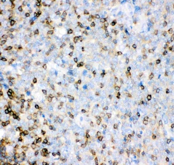
IHC(P) analysis of Human Tonsil Tissue using Anti-IL-16 antibody.
Anti-IL-16 antibody, IHC(P): Human Tonsil Tissue.
WB analysis of Recombinant Human IL-16 Protein 0.5ng using Anti-IL-16 antibody.
Anti-IL-16 antibody, Western blotting. All lanes: Anti IL-16 at 0.5 ug/ml. WB: Recombinant Human IL-16 Protein 0.5 ng. Predicted bind size: 17 KD. Observed bind size: 17 KD.
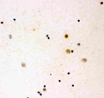
IHC analysis of IL16 using anti-IL16 antibody. IL16 was detected in immunocytochemical section of human cord blood. Enzyme antigen retrieval was performed using IHC enzyme antigen retrieval reagent for 15 mins. The cells were blocked with 10% goat serum. And then incubated with 1 µg/ml rabbit anti-IL16 Antibody overnight at 4C. Biotinylated goat anti-rabbit IgG was used as secondary antibody and incubated for 30 minutes at 37C. The section was developed using Strepavidin-Biotin-Complex (SABC) with DAB as the chromogen.
Western blot analysis of IL16 using anti-IL16 antibody. Electrophoresis was performed on a 5-20% SDS-PAGE gel at 70V (Stacking gel) / 90V (Resolving gel) for 2-3 hours. The sample well of each lane was loaded with 50 ug of sample under reducing conditions. Lane 1: human U937 whole cell lysates, After Electrophoresis, proteins were transferred to a Nitrocellulose membrane at 150mA for 50-90 minutes. Blocked the membrane with 5% Non-fat Milk/ TBS for 1.5 hour at RT. The membrane was incubated with rabbit anti-IL16 antigen affinity purified polyclonal antibody at 0.5 µg/mL overnight at 4C, then washed with TBS-0.1% Tween 3 times with 5 minutes each and probed with a goat anti-rabbit IgG-HRP secondary antibody at a dilution of 1:10000 for 1.5 hour at RT. The signal is developed using an Enhanced Chemiluminescent detection (ECL) kit with Tanon 5200 system. A specific band was detected for IL16 at approximately 45-55 KD. The expected band size for IL16 is at 142 KD.
* VAT and and shipping costs not included. Errors and price changes excepted