E.coli-derived human IKK beta recombinant protein (Position: E398-S756). Human IKK beta shares 91% and 90% amino acid (aa) sequences identity with mouse and rat IKK beta, respectively.
Each vial contains antibody formulated with stabilizing components, 0.9 mg NaCl, 0.2 mg Na2HPO4, and 0.05 mg NaN3. *This antibody is supplied in a stabilized formulation. Compatibility with conjugation reactions depends on the chemistry of the conjugation
Form:
Lyophilized
Target:
Inhibitor of nuclear factor kappa-B kinase subunit beta
Application Dilute:
Western blot, 0.1-0.5µg/ml, Human Immunohistochemistry (Paraffin-embedded Section), 0.5-1µg/ml, Human Immunocytochemistry/Immunofluorescence, 2µg/ml, Human Flow Cytometry (Fixed), 1-3µg/1x10 6 cells, Human
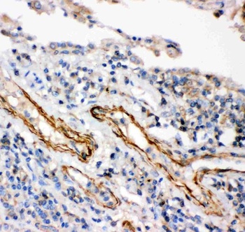
IHC(P) analysis of Human Lung Cancer Tissue using Anti-IKK beta antibody.
Anti-IKK beta antibody, Western blotting. All lanes: Anti IKK beta at 0.5 ug/ml. Lane 1: HEPG2 Whole Cell Lysate at 40 ug. Lane 2: COLO320 Whole Cell Lysate at 40 ug. Lane 3: M231 Whole Cell Lysate at 40 ug. Lane 4: HT1080 Whole Cell Lysate at 40 ug. Predicted bind size: 87 KD. Observed bind size: 87 KD.
Anti-IKK beta antibody, Western blotting. All lanes: Anti IKK beta at 0.5 ug/ml. WB: Recombinant Human IKK beta Protein 0.5 ng. Predicted bind size: 43 KD. Observed bind size: 43 KD.
Anti-IKK beta antibody, IHC(P): Human Lung Cancer Tissue.
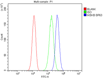
Flow Cytometry analysis of 293T cells using anti-IKK beta antibody. Overlay histogram showing 293T cells (Blue line). To facilitate intracellular staining, cells were fixed with 4% paraformaldehyde and permeabilized with permeabilization buffer. The cells were blocked with 10% normal goat serum. And then incubated with rabbit anti-IKK beta Antibody (1 µg/1x10 6 cells) for 30 min at 20C. DyLight488 conjugated goat anti-rabbit IgG (5-10 µg/1x10 6 cells) was used as secondary antibody for 30 minutes at 20C. Isotype control antibody (Green line) was rabbit IgG (1 µg/1x10 6) used under the same conditions. Unlabelled sample without incubation with primary antibody and secondary antibody (Red line) was used as a blank control.
IF analysis of IKK beta using anti-IKK beta antibody. IKK beta was detected in immunocytochemical section of A431 cells. Enzyme antigen retrieval was performed using IHC enzyme antigen retrieval reagent for 15 mins. The cells were blocked with 10% goat serum. And then incubated with 2 µg/mL rabbit anti-IKK beta Antibody overnight at 4C. DyLight594 Conjugated Goat Anti-Rabbit IgG was used as secondary antibody at 1:100 dilution and incubated for 30 minutes at 37C. The section was counterstained with DAPI. Visualize using a fluorescence microscope and filter sets appropriate for the label used.
* VAT and and shipping costs not included. Errors and price changes excepted