Purified polyclonal antibody supplied in PBS with 0.09% (W/V) sodium azide. This antibody is purified through a protein A column, followed by peptide affinity purification.
Application Dilute:
IHC-P - 1:100-500, IF - 1:25, WB - 1:1000, FC - 1:10-50
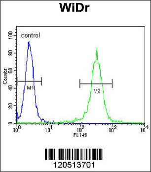
ATP5B Antibody (Center) flow cytometric analysis of WiDr cells (right histogram) compared to a negative control cell (left histogram). FITC-conjugated goat-anti-rabbit secondary antibodies were used for the analysis.
Immunohistochemical analysis of paraffin-embedded H. liver section using ATP5B Antibody (Center). Diluted at 1:25 dilution. A undiluted biotinylated goat polyvalent antibody was used as the secondary, followed by DAB staining.
Immunohistochemical analysis of paraffin-embedded H. small intestine section using ATP5B Antibody (Center). Diluted at 1:25 dilution. A undiluted biotinylated goat polyvalent antibody was used as the secondary, followed by DAB staining.
Fluorescent image of SK-BR-3 cells stained with ATP5B Antibody (Center). Diluted at 1:25 dilution. An Alexa Fluor 488-conjugated goat anti-rabbit lgG at 1:400 dilution was used as the secondary antibody (green). DAPI was used to stain the cell nuclear (blue).
Formalin-fixed and paraffin-embedded human brain tissue reacted with ATP5B Antibody (Center), which was peroxidase-conjugated to the secondary antibody, followed by DAB staining. This data demonstrates the use of this antibody for immunohistochemistry, clinical relevance has not been evaluated.
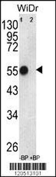
Western blot analysis of ATP5B Antibody (Center) Pab pre-incubated without (lane 1) and with (lane 2) blocking peptide in WiDr cell line lysate. ATP5B (arrow) was detected using the purified Pab.
Western blot analysis of ATP5B (arrow) using rabbit polyclonal ATP5B Antibody (Center). 293 cell lysates (2 ug/lane) either nontransfected (Lane 1) or transiently transfected with the ATP5B gene (Lane 2).
* VAT and and shipping costs not included. Errors and price changes excepted