Purified polyclonal antibody supplied in PBS with 0.09% (W/V) sodium azide. This antibody is purified through a protein A column, followed by peptide affinity purification.
Application Dilute:
WB - 1:1000, IHC-P - 1:100-500
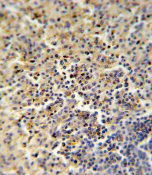
FOLR2 Antibody (N-term) IHC analysis in formalin fixed and paraffin embedded human spleen followed by peroxidase conjugation of the secondary antibody and DAB staining. This data demonstrates the use of the FOLR2 Antibody (N-term) for immunohistochemistry. Clinical relevance has not been evaluated.
Western blot analysis of lysate from human placenta tissue, using FOLR2 Antibody (N-term). Diluted at 1:1000. A goat anti-rabbit IgG H&L (HRP) at 1:10000 dilution was used as the secondary antibody. Lysate at 20 ug.
Western blot analysis of lysate from mouse heart tissue lysate, using FOLR2 Antibody (N-term). Diluted at 1:1000. A goat anti-rabbit IgG H&L (HRP) at 1:5000 dilution was used as the secondary antibody. Lysate at 35 ug.
Western blot analysis of lysate from human placenta tissue lysate, using FOLR2 Antibody (N-term). Diluted at 1:1000. A goat anti-rabbit IgG H&L (HRP) at 1:5000 dilution was used as the secondary antibody. Lysate at 35 ug.
Western blot analysis of lysate from human placenta tissue lysate, using FOLR2 Antibody (N-term). Diluted at 1:1000. A goat anti-rabbit IgG H&L (HRP) at 1:10000 dilution was used as the secondary antibody. Lysate at 35 ug.
Western blot analysis of lysates from KG-1, MCF-7, MDA-MB-453 cell line (from left to right), using FOLR2 Antibody (N-term). Diluted at 1:1000 at each lane. A goat anti-rabbit IgG H&L (HRP) at 1:5000 dilution was used as the secondary antibody. Lysates at 35 ug per lane.
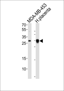
Western blot analysis of lysates from MDA-MB-453 cell line, human placenta tissue lysate (from left to right), using FOLR2 Antibody (N-term). Diluted at 1:1000 at each lane. A goat anti-rabbit IgG H&L (HRP) at 1:10000 dilution was used as the secondary antibody. Lysates at 20 ug per lane.
* VAT and and shipping costs not included. Errors and price changes excepted