Purified polyclonal antibody supplied in PBS with 0.05% (V/V) Proclin 300. This antibody is purified through a protein A column, followed by peptide affinity purification.
Application Dilute:
WB - 1:2000, IHC-P-Leica - 1:500
S100B Antibody flow cytometric analysis of A375 cells (right histogram) compared to a negative control cell (left histogram). FITC-conjugated goat-anti-rabbit secondary antibodies were used for the analysis.
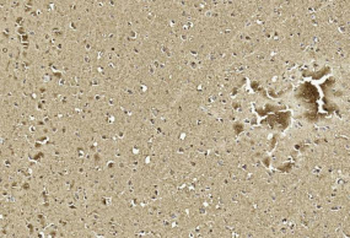
Immunohistochemical analysis of paraffin-embedded Human brain section using Pink1. Diluted at 1:1000 dilution. A undiluted biotinylated goat polyvalent antibody was used as the secondary, followed by DAB staining.
Immunohistochemical analysis of paraffin-embedded Human breast tissue was performed on the Leica BOND RXm. Tissue was fixed with formaldehyde at room temperature, antigen retrieval was by heat mediation with a EDTA buffer (pH9.0). Samples were incubated with primary antibody (1:500) for 1 hours at room temperature. A undiluted biotinylated CRF Anti-Polyvalent HRP Polymer antibody was used as the secondary antibody.
Immunohistochemical analysis of paraffin-embedded Human melanoma tissue was performed on the Leica BOND RXm. Tissue was fixed with formaldehyde at room temperature, antigen retrieval was by heat mediation with a EDTA buffer (pH9.0). Samples were incubated with primary antibody (1:500) for 1 hours at room temperature. A undiluted biotinylated CRF Anti-Polyvalent HRP Polymer antibody was used as the secondary antibody.
Anti-S100B Antibody at 1:2000 dilution + Human brain whole tissue lysate. Lysates/proteins at 20 µg per lane. Secondary Goat Anti-Rabbit IgG, (H+L), Peroxidase conjugated at 1/10000 dilution. Predicted band size: 11 kDa. Blocking/Dilution buffer: 5% NFDM/TBST.
Western blot analysis of lysates from A431 cell line, mouse brain, rat brain tissue (from left to right), using S100B Antibody. Diluted at 1:1000 at each lane. A goat anti-rabbit IgG H&L (HRP) at 1:10000 dilution was used as the secondary antibody. Lysates at 20 ug per lane.
All lanes: Anti-S100B Antibody at 1:2000 dilution. Lane 1: Human brain lysate. Lane 2: C2C12 whole cell lysate. Lane 3: Rat brain lysate. Lysates/proteins at 20 µg per lane. Secondary Goat Anti-Rabbit IgG, (H+L), Peroxidase conjugated at 1/10000 dilution. Predicted band size: 11 kDa. Blocking/Dilution buffer: 5% NFDM/TBST.
* VAT and and shipping costs not included. Errors and price changes excepted