Purified polyclonal antibody supplied in PBS with 0.05% (V/V) Proclin 300. This antibody is prepared by Saturated Ammonium Sulfate (SAS) precipitation followed by dialysis against PBS.
Application Dilute:
WB - 1:1000
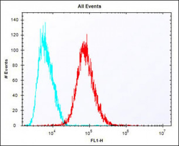
Overlay histogram showing HepG2 cells stained (red line). The cells were fixed with 2% paraformaldehyde (10 min) and then permeabilized with 90% methanol for 10 min. The cells were then icubated in 2% bovine serum albumin to block non-specific protein-protein interactions followed by the antibody (1:25 dilution) for 60 min at 37C. The secondary antibody used was Alexa Fluor 488 goat anti-rabbit lgG (H+L) at 1/400 dilution for 40 min at 37C. Isotype control antibody (blue line) was rabbit IgG1 (1 µg/1x10 6 cells) used under the same conditions. Acquisition of > 10000 events was performed.
IDUA Antibody (Center) immunohistochemistry analysis in formalin fixed and paraffin embedded human prostate carcinoma followed by peroxidase conjugation of the secondary antibody and DAB staining. This data demonstrates the use of the IDUA Antibody (Center) for immunohistochemistry. Clinical relevance has not been evaluated.
IDUA Antibody (Center) western blot analysis in Hela cell line lysates (35 ug/lane). This demonstrates the IDUA antibody detected the IDUA protein (arrow).
Anti-IDUA Antibody (Center) at 1:1000 dilution + human lung lysate. Lysates/proteins at 20 µg per lane. Secondary Goat Anti-Rabbit IgG, (H+L), Peroxidase conjugated at 1/10000 dilution. Predicted band size: 73 kDa. Blocking/Dilution buffer: 5% NFDM/TBST.
Anti-IDUA Antibody (Center) at 1:2000 dilution + human liver lysates. Lysates/proteins at 20 µg per lane. Secondary Goat Anti-Rabbit IgG, (H+L), Peroxidase conjugated at 1/10000 dilution. Predicted band size: 73 kDa. Blocking/Dilution buffer: 5% NFDM/TBST.
Western blot analysis of lysates from A549 cell line, rat lung tissue lysate (from left to right), using IDUA Antibody (Center). Diluted at 1:1000 at each lane. A goat anti-rabbit IgG H&L (HRP) at 1:10000 dilution was used as the secondary antibody. Lysates at 35 ug per lane.
All lanes: Anti-IDUA Antibody (Center) at 1:2000 dilution. Lane 1: human liver lysates. Lane 2: human lung lysates. Lysates/proteins at 20 µg per lane. Secondary Goat Anti-Rabbit IgG, (H+L), Peroxidase conjugated at 1/10000 dilution. Predicted band size: 73 kDa. Blocking/Dilution buffer: 5% NFDM/TBST.
* VAT and and shipping costs not included. Errors and price changes excepted