Purified polyclonal antibody supplied in PBS with 0.05% (V/V) Proclin 300. This antibody is purified through a protein A column, followed by peptide affinity purification.
Application Dilute:
WB - 1:500
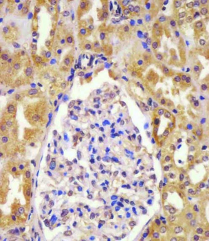
Staining VHL in human kidney tissue sections by Immunohistochemistry (IHC-P - paraformaldehyde-fixed, paraffin-embedded sections). Tissue was fixed with formaldehyde and blocked with 3% BSA for 0.5 hour at room temperature, antigen retrieval was by heat mediation with a citrate buffer (pH6). Samples were incubated with primary antibody (1/25) for 1 hours at 37C. A undiluted biotinylated goat polyvalent antibody was used as the secondary antibody.
Staining VHL in human pancreas tissue sections by Immunohistochemistry (IHC-P - paraformaldehyde-fixed, paraffin-embedded sections). Tissue was fixed with formaldehyde and blocked with 3% BSA for 0.5 hour at room temperature, antigen retrieval was by heat mediation with a citrate buffer (pH6). Samples were incubated with primary antibody (1/25) for 1 hours at 37C. A undiluted biotinylated goat polyvalent antibody was used as the secondary antibody.
Immunofluorescent analysis of 4% paraformaldehyde-fixed, 0.1% Triton X-100 permeabilized HeLa (human cervical epithelial adenocarcinoma cell line) cells labeling VHL at 1/25 dilution, followed by Dylight 488-conjugated goat anti-rabbit IgG secondary antibody at 1/200 dilution (green). Immunofluorescence image showing nucleus and cytoplasm staining on HeLa cell line. Cytoplasmic actin is detected with Dylight 554 Phalloidin at 1/100 dilution (red). The nuclear counter stain is DAPI (blue).
Western blot analysis of VHL antibody (N-term) in HepG2 cell line lysates (35 ug/lane). VHL (arrow) was detected using the purified Pab.
Anti-VHL Antibody (N-term) at 1:2000 dilution + F9 whole cell lysate. Lysates/proteins at 20 µg per lane. Secondary Goat Anti-Rabbit IgG, (H+L), Peroxidase conjugated at 1/10000 dilution. Predicted band size: 24 kDa. Blocking/Dilution buffer: 5% NFDM/TBST.
All lanes: Anti-VHL Antibody (N-term) at 1:500 dilution. Lane 1: F9 whole cell lysate. Lane 2: Mouse testis lysate. Lysates/proteins at 20 µg per lane. Secondary Goat Anti-Rabbit IgG, (H+L), Peroxidase conjugated at 1/10000 dilution. Predicted band size: 24 kDa. Blocking/Dilution buffer: 5% NFDM/TBST.
All lanes: Anti-VHL Antibody (N-term) at 1:1000 dilution. Lane 1: F9 whole cell lysate. Lane 2: Mouse testis lysate. Lysates/proteins at 20 µg per lane. Secondary Goat Anti-Rabbit IgG, (H+L), Peroxidase conjugated at 1/10000 dilution. Predicted band size: 24 kDa. Blocking/Dilution buffer: 5% NFDM/TBST.
* VAT and and shipping costs not included. Errors and price changes excepted