anti FTCD antibody, anti LCHC1 antibody, anti formiminotransferase cyclodeaminase antibody, anti formimidoyltransferase cyclodeaminase antibody, anti LC1 autoantigen antibody
Goat polyclonal antibody to FTCD
Clonality:
Polyclonal
Molecular Weight:
58.9, 58.9, 61.3
Buffer:
Supplied at 0.5 mg/ml in Tris saline, 0.02% sodium azide, pH 7.3 with 0.5% bovine serum albumin. Aliquot and store at -20C. Minimize freezing and thawing.
Sequence:
CLREQGRGKDQPGRL
Target:
58KGolgi protein(Internal)/FTCD
Application Dilute:
ELISA: 1:64000, WB: 0.1-0.3 µg/ml, IHC-P: 10ug/ml
Application Notes:
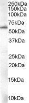
Application Notes: ELISA: Peptide ELISA: antibody detection limit dilution 1:64000.WB: Approx 60kDa band observed in Human Liver lysates (calculated MW of 58.9kDa according to NP_006648.1and NP_996848.1). Recommended concentration: 0.01-0.03 µg/ml
Western blot analysis of Human Liver lysate using FTCD antibody
Immunohistochemical staining of Human Liver using FTCD antibody
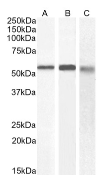
0.75 µg/ml staining of Human (A) and Mouse (B) and (0.5 ug/ml) of Rat Liver lysate (35 µg protein in RIPA buffer). Detected by chemiluminescence.
Immunofluorescence analysis of paraformaldehyde fixed Caco-2 cells, permeabilized with 0.15% Triton. Primary incubation 1hr (10 ug/ml) followed by Alexa Fluor 488 secondary antibody (2 ug/ml), showing membrane and cytoplasmic staining. The nuclear stain is DAPI (blue). Negative control: Unimmunized goat IgG (10 ug/ml) followed by Alexa Fluor 488 secondary antibody (2 ug/ml).
6 µg/ml staining of paraffin embedded Human Liver. Heat induced antigen retrieval with citrate buffer pH6, HRP-staining.
Negative Control showing staining of paraffin embedded Human Liver, with no primary antibody.
* VAT and and shipping costs not included. Errors and price changes excepted