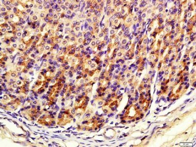
Immunohistochemical staining of Rat stomach tissue using FITM1 antibody.
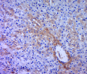
Immunohistochemical staining of rat liver tissue using FITM1 antibody.
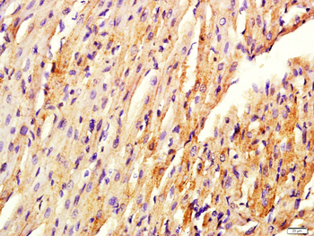
Paraformaldehyde-fixed, paraffin embedded (Rat heart), Antigen retrieval by boiling in sodium citrate buffer (pH6.0) for 15 min, Block endogenous peroxidase by 3% hydrogen peroxide for 20 minutes, Blocking buffer (normal goat serum) at 37C for 30 min, Antibody incubation with (Fat inducing transcript, FITM1) Polyclonal Antibody, Unconjugated (orb183695) at 1:400 overnight at 4C, followed by a conjugated secondary antibody for 20 minutes and DAB staining.
Paraformaldehyde-fixed, paraffin embedded (rat liver tissue), Antigen retrieval by boiling in sodium citrate buffer (pH6.0) for 15 min, Block endogenous peroxidase by 3% hydrogen peroxide for 20 minutes, Blocking buffer (normal goat serum) at 37C for 30 min, Antibody incubation with (FITM1) Polyclonal Antibody, Unconjugated (orb183695) at 1:400 overnight at 4C, followed by a conjugated secondary for 20 minutes and DAB staining.
Paraformaldehyde-fixed, paraffin embedded (Rat stomach), Antigen retrieval by boiling in sodium citrate buffer (pH6.0) for 15 min, Block endogenous peroxidase by 3% hydrogen peroxide for 20 minutes, Blocking buffer (normal goat serum) at 37C for 30 min, Antibody incubation with (Fat inducing transcript, FITM1) Polyclonal Antibody, Unconjugated (orb183695) at 1:400 overnight at 4C, followed by a conjugated secondary antibody for 20 minutes and DAB staining.
Protein: 293T (human) lysate at 40 ug, Primary: rabbit Anti-FITM1 (orb183695) at 1:300, Secondary: HRP conjugated Goat-Anti-rabbit IgG (orb572747) at 1:5000, Predicted band size: 32 kD, Observed band size: 28 kD.
* VAT and and shipping costs not included. Errors and price changes excepted