anti SIAH1 antibody, anti seven in absentia homolog 1 (Drosophila) antibody, anti Siah-1 antibody, anti hSIAH1 antibody, anti HUMSIAH antibody, anti Siah-1a antibody, anti FLJ08065 antibody, anti seven in absentia homolog 1 antibody, anti sonic hedgehog homolog antibody
Goat polyclonal antibody to SIAH1
Clonality:
Polyclonal
Molecular Weight:
31.1, 34.6
Buffer:
Supplied at 0.5 mg/ml in Tris saline, 0.02% sodium azide, pH 7.3 with 0.5% bovine serum albumin. Aliquot and store at -20C. Minimize freezing and thawing.
Sequence:
SRQTATALPTGTSKC
Target:
SIAH1
Application Dilute:
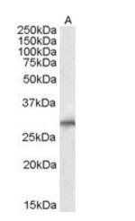
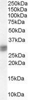
Peptide ELISA: antibody detection limit dilution 1:128000. Western blot: Approx. 37kDa band observed in Human Liver lysates and approx. 30+37kDa bands in Rat Liver lysates (calculated MW of 34.6kDa according to Human NP_001006611.1 and 31.1kDa according t
Western blot analysis of Human Liver lysate using SIAH1 antibody
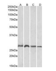
Western blot analysis of Mouse and Rat Brain and Liver lysates using SIAH1 antibody
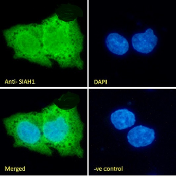
Immunofluorescence analysis of paraformaldehyde fixed HepG2 cells, permeabilized with 0.15% Triton. Primary incubation 1hr (10 ug/ml) followed by Alexa Fluor 488 secondary antibody (2 ug/ml), showing nuclear and cytoplasmic staining. The nuclear stain is DAPI (blue). Negative control: Unimmunized goat IgG (10 ug/ml) followed by Alexa Fluor 488 secondary antibody
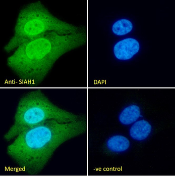
Immunofluorescence analysis of paraformaldehyde fixed U2OS cells, permeabilized with 0.15% Triton. Primary incubation 1hr (10 ug/ml) followed by Alexa Fluor 488 secondary antibody (2 ug/ml), showing nuclear and cytoplasmic staining. The nuclear stain is DAPI (blue). Negative control: Unimmunized goat IgG (10 ug/ml) followed by Alexa Fluor 488 secondary antibody.
Primary incubation 1 hour at room temperature. Images A+B: Human Liver lysate + peptide, incubation at primary Ab concentration 1 ug/ml, C+D: Rat Liver lysate + peptide, incubation at primary Ab concentration 3 ug/ml. (Loaded 35 µg protein in RIPA buffer, per lane). Detected by chemiluminescence.
Western blot analysis of Human Liver lysate using SIAH1 antibody
* VAT and and shipping costs not included. Errors and price changes excepted