anti TRPM7 antibody, anti transient receptor potential cation channel, subfamily M, member 7 antibody, anti CHAK antibody, anti CHAK1 antibody, anti LTRPC7 antibody, anti FLJ20117 antibody, anti TRP-PLIK antibody, anti LTRPC ion channel family member 7 antibody, anti homolog of mouse transient receptor potential-phospholipase C-interacting kinase CHaK hypothetical protein FLJ20117 antibody, anti FLJ25718 antibody
Goat polyclonal antibody to TRPM7
Clonality:
Polyclonal
Molecular Weight:
213, 213
Buffer:
Supplied at 0.5 mg/ml in Tris saline, 0.02% sodium azide, pH 7.3 with 0.5% bovine serum albumin. Aliquot and store at -20C. Minimize freezing and thawing.
Sequence:
TKESESTNSVRLML
Target:
TRPM7 / LTRPC7
Application Dilute:
ELISA: 1:8000, IHC-P: 4-6 µg/ml
Application Notes:
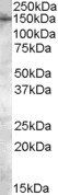
Application Notes: ELISA: Peptide ELISA: antibody detection limit dilution 1:16000.WB: Approx. 150kDa band observed in Human Brain (Cerebellum) lysates (calculated MW of 213kDa according to NP_060142.2). The observed molecular weight corresponds to earlier findings in literature with different antibodies (Clark et al PLoS ONE. 2008 Mar 26,3(3):e1876., PMID: 18365021). Recommended concentration: 0.3- 1 µg/ml
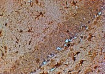
Immunohistochemical staining of Mouse Hippocampus using TRPM7 antibody
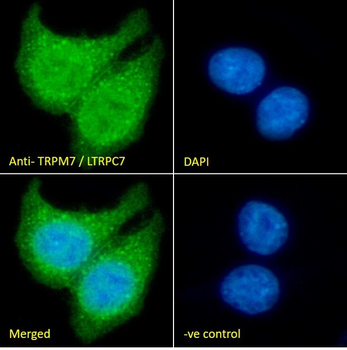
Immunofluorescence analysis of paraformaldehyde fixed MCF7 cells, permeabilized with 0.15% Triton. Primary incubation 1hr (10 ug/ml) followed by Alexa Fluor 488 secondary antibody (2 ug/ml), showing nuclear and cytoplasmic staining. The nuclear stain is DAPI (blue). Negative control: Unimmunized goat IgG (10 ug/ml) followed by Alexa Fluor 488 secondary antibody (2 ug/ml).
6 µg/ml staining of paraffin embedded Human Kidney. Heat induced antigen retrieval with citrate buffer pH6, HRP-staining.
Negative Control showing staining of paraffin embedded Human Kidney, with no primary antibody.
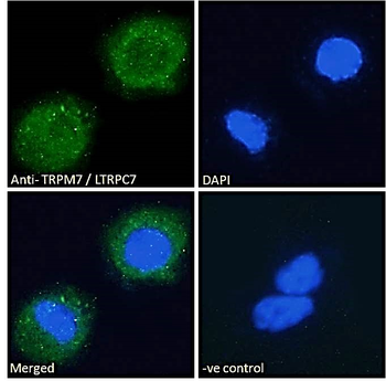
Immunofluorescence analysis of paraformaldehyde fixed A431 cells, permeabilized with 0.15% Triton. Primary incubation 1hr (10 ug/ml) followed by Alexa Fluor 488 secondary antibody (2 ug/ml), showing vesicle staining. The nuclear stain is DAPI (blue). Negative control: Unimmunized goat IgG (10 ug/ml) followed by Alexa Fluor 488 secondary antibody (2 ug/ml).
Western blot analysis of human cerebellum lysate using TRPM7 antibody
* VAT and and shipping costs not included. Errors and price changes excepted