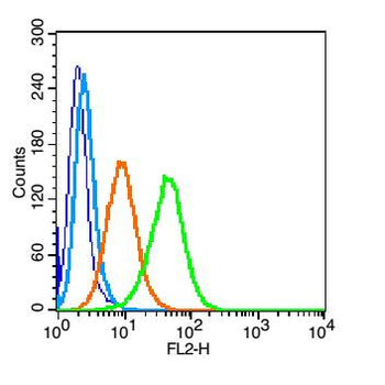
Flow cytometric analysis of U937 cell using CD101 antibody.
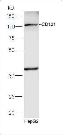
Western blot analysis of HepG2 lysates using CD101 antibody.
Blank control (blue): U937 (fixed with 2% paraformaldehyde (10 min)). Primary Antibody: Rabbit Anti-CD101 antibody (orb182947), dilution: 1 µg in 100 µl 1X PBS containing 0.5% BSA, Isotype Control Antibody: Rabbit IgG (orange), used under the same conditions, Secondary Antibody: Goat anti-rabbit IgG-PE (white blue), dilution: 1:200 in 1X PBS containing 0.5% BSA.
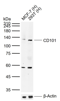
Sample: Lane 1: Human MCF-7 cell lysates, Lane 2: Human 293T cell lysates, Primary: Anti-CD101 (orb182947) at 1/1000 dilution, Secondary: IRDye800CW Goat Anti-Rabbit IgG at 1/20000 dilution, Predicted band size: 113 kDa, Observed band size: 125 kDa.
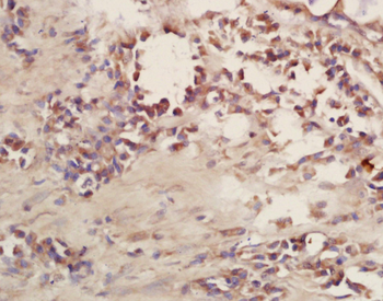
Tissue/Cell: human bladder carcinoma, 4% Paraformaldehyde-fixed and paraffin-embedded, Antigen retrieval: citrate buffer (0.01M, pH 6.0), Boiling bathing for 15 min, Block endogenous peroxidase by 3% Hydrogen peroxide for 30 min, Blocking buffer (normal goat serum) at 37C for 20 min, Incubation: Anti-CD101 Polyclonal Antibody, Unconjugated (orb182947) 1:200, overnight at 4C, followed by conjugation to the secondary antibody and DAB staining.
Tissue/Cell: human lung carcinoma, 4% Paraformaldehyde-fixed and paraffin-embedded, Antigen retrieval: citrate buffer (0.01M, pH 6.0), Boiling bathing for 15 min, Block endogenous peroxidase by 3% Hydrogen peroxide for 30 min, Blocking buffer (normal goat serum) at 37C for 20 min, Incubation: Anti-CD101 Polyclonal Antibody, Unconjugated (orb182947) 1:200, overnight at 4C, followed by conjugation to the secondary antibody and DAB staining.
* VAT and and shipping costs not included. Errors and price changes excepted