TIGIT Antibody is supplied in PBS containing 0.02% sodium azide and 50% glycerol.
Form:
Liquid
Target:
TIGIT
Application Notes:
Application Notes: TIGIT antibody can be used for immunohistochemistry starting at 2 µg/mL. For immunofluorescence start at 1 µg/mL. For flow cytometry at 1 µg/ml. For immunocytochemistry at 1 µg/mL. For Western blot at 1 µg/mL. Antibody validated: Western Blot in human samples, Immunohistochemistry in human samples, Immunocytochemistry in human samples, Immunofluorescence in human samples and Flow Cytometry in human samples. All other applications and species not yet tested
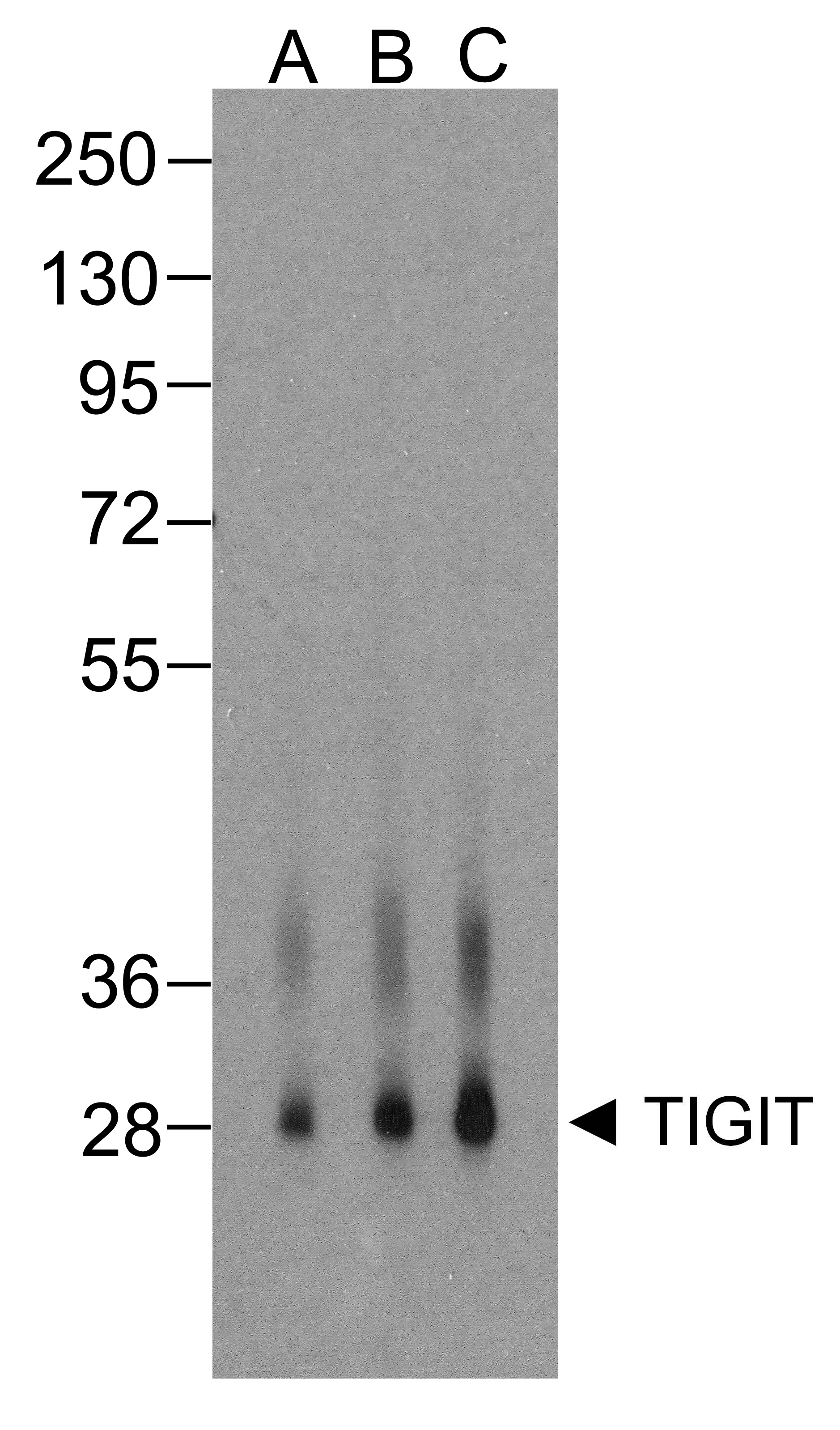
Western blot analysis of TIGIT in over expressing HEK293 cells using orb1240140 antibody at (A) 0.25 µg/ml, (B) 0.5 µg/ml, and (C) 1 µg/ml.
Immunocytochemistry of TIGIT in over expressing HEK293 cells using TIGIT antibody and control mouse IgG antibody (left corner box) at 1 µg/ml.
Immunofluorescence of TIGIT in over expressing HEK293 cells using TIGIT Antibody at 1 µg/ml. Green: TIGIT Antibody [4A11] (orb1240140) Blue: DAPI staining
Immunofluorescence of TIGIT in human stomach carcinoma tissue using TIGIT Antibody at 5 µg/ml. Green: TIGIT Antibody [4A11] (orb1240140) Blue: DAPI staining
Immunohistochemistry of TIGIT in human stomach carcinoma tissue using TIGIT Antibody and control mouse IgG (corner box) at 2 µg/ml.
Flow cytometry analysis of TIGIT over expressing HEK293 cells using TIGIT antibody at 1 µg/ml. Blue: untransfected HEK293 cells. Yellow: TIGIT over expressing HEK293 cells.
Titration curve analysis of TIGIT mAbs to detect recombinant TIGIT in ELISA with orb1240147, orb1240136, orb1240137, orb1240140, orb1240146 and orb1240115 antibodies at decreasing concentrations.
A sandwich ELISA was performed using the anti-TIGIT mAb orb1240140 (5 µg/ml) as the capture antibody. Biotin-labeled anti-TIGIT mAb orb1238175 (1 µg/ml) and streptavidin-HRP (0.1 µg/ml) were used for detection. Detection range is from 10 ng to 40 pg.
* VAT and and shipping costs not included. Errors and price changes excepted