Anti-TET1 antibody (orb1240105) was raised against an 18 amino acid peptide near the carboxy terminus of human TET1. The immunogen is located within amino acids 2030 - 2080 of TET1.
TET1 Antibody is supplied in PBS containing 0.02% sodium azide.
Form:
Liquid
Target:
TET1
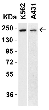
WB Validation of TET1 in Human Cell Lines. Loading: 15 µg of lysate Antibodies: TET1 orb1240105, 1 µg/mL, 1 h incubation at RT in 5% NFDM/TBST. Secondary: Goat Anti-Rabbit IgG HRP conjugate at 1:10000 dilution.
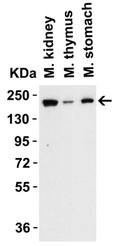
WB Validation of TET1 in Mouse Tissues. Loading: 15 µg of lysate Antibodies: TET1 orb1240105, 1 µg/mL, 1 h incubation at RT in 5% NFDM/TBST. Secondary: Goat Anti-Rabbit IgG HRP conjugate at 1:10000 dilution.
WB Validation of TET1 in Rat Tissues. Loading: 15 µg of lysate Antibodies: TET1 orb1240105, 1 µg/mL, 1 h incubation at RT in 5% NFDM/TBST. Secondary: Goat Anti-Rabbit IgG HRP conjugate at 1:10000 dilution.
Immunofluorescence Validation of TET1 in HeLa Cells. Immunofluorescent analysis of 4% paraformaldehyde-fixed Hela cells labeling TET1 with orb1240105 at 20 µg/mL, followed by goat anti-rabbit IgG secondary antibody at 1/500 dilution (green) and DAPI antibody (blue).
Immunofluorescence Validation of TET1 in Mouse Brain Tissue. Immunofluorescent analysis of 4% paraformaldehyde-fixed mouse brain tissue labeling TET1 with orb1240105 at 20 µg/mL, followed by goat anti-rabbit IgG secondary antibody at 1/500 dilution (red) and DAPI antibody (blue).
Immunohistochemistry Validation of TET1 in Human Testis Tissue. Immunohistochemical analysis of paraffin-embedded human testis tissue using anti-TET1 antibody (orb1240105) at 1 µg/ml. Tissue was fixed with formaldehyde and blocked with 10% serum for 1 h at RT, antigen retrieval was by heat mediation with a citrate buffer (pH6). Samples were incubated with primary antibody overnight at 4C. A goat anti-rabbit IgG H&L (HRP) at 1/250 was used as secondary. Counter stained with Hematoxylin.
Immunohistochemistry Validation of TET1 in Mouse Kidney Tissue. Immunohistochemical analysis of paraffin-embedded mouse kidney tissue using anti-TET1 antibody (orb1240105) at 1 µg /ml. Tissue was fixed with formaldehyde and blocked with 10% serum for 1 h at RT, antigen retrieval was by heat mediation with a citrate buffer (pH6). Samples were incubated with primary antibody overnight at 4C. A goat anti-rabbit IgG H&L (HRP) at 1/250 was used as secondary. Counter stained with Hematoxylin.
Immunohistochemistry Validation of TET1 in Rat Liver Tissue. Immunohistochemical analysis of paraffin-embedded rat liver tissue using anti-TET1 antibody (orb1240105) at 1 µg /ml. Tissue was fixed with formaldehyde and blocked with 10% serum for 1 h at RT, antigen retrieval was by heat mediation with a citrate buffer (pH6). Samples were incubated with primary antibody overnight at 4C. A goat anti-rabbit IgG H&L (HRP) at 1/250 was used as secondary. Counter stained with Hematoxylin.
* VAT and and shipping costs not included. Errors and price changes excepted