ST2 antibody was raised against a synthetic peptide corresponding to 16 amino acids at the amino-terminus of mouse ST2.This peptide is common to all three known ST2 isoforms.The immunogen is located within amino acids 40 - 90 of ST2.
ST2 Antibody is supplied in PBS containing 0.02% sodium azide.
Form:
Liquid
Target:
Il1rl1
Application Notes:
Application Notes: WB: 1-2 µg/mL, IHC: 1-5 µg/mL, IF: 5-20 µg/mL. Antibody validated: Western Blot in human, mouse and rat samples, Immunocytochemistry and Immunofluorescence in human, mouse and rat samples. All other applications and species not yet tested
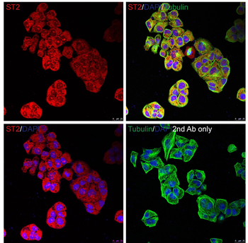
Immunoflouorescence Validation of ST2 in HeLa Cells. Immunofluorescent analysis of PFA-fixed HeLa cells labeling ST2 with orb1240035 at 20 µg/mL, followed by goat anti-rabbit IgG secondary antibody at 1/1000 dilution (red) and DAPI staining (blue). Alpha tubulin was stained with anti-alpha tubulin antibody following by goat anti-mouse IgG secondary antibody (green). Images were captured with confocal microscopy.
WB Validation in Human, Mouse, and Rat Cell Lines. Loading: 15 µg of lysate per lane Antibodies: ST2 orb1240035, 1 µg/mL, 1 h incubation at RT in 5% NFDM/TBST. Secondary: Goat Anti-Rabbit IgG HRP conjugate at 1:10000 dilution.
Western Blot Validation in 293 Cells. Antibodies: ST2 orb1240035, 1 µg/mL, 1 h incubation at RT in 5% NFDM/TBST. Secondary: Goat anti-rabbit IgG HRP conjugate at 1:10000 dilution. Lane A: In the absence of blocking peptide Lane B: In the presence of blocking peptide.
Western Blot Validation in Mouse Heart. Loading: 15 µg of lysate Antibodies: ST2 orb1240035, 1 µg/mL, 1 h incubation at RT in 5% NFDM/TBST. Secondary: Goat anti-rabbit IgG HRP conjugate at 1:10000 dilution.
Immunofluorescence Validation of ST2 in Human Lung. Immunofluorescent analysis of PFA-fixed human lung tissue labeling ST2 with orb1240035 at 10 µg/mL, followed by goat anti-rabbit IgG secondary antibody at 1/1000 dilution (red) and DAPI staining (blue).
Immunofluorescence Validation of ST2 in Human Lung Cancer Tissue. Immunofluorescent analysis of 4% paraformaldehyde-fixed human lung cancer tissue labeling ST2 with orb1240035 at 5 µg/mL, followed by goat anti-rabbit IgG secondary antibody at 1/500 dilution (green) DAPI staining (blue).
Immunofluorescence Validation of ST2 in Mouse Kidney. Immunofluorescent analysis of PFA-fixed mouse kidney tissue labeling ST2 with orb1240035 at 10 µg/mL, followed by goat anti-rabbit IgG secondary antibody at 1/1000 dilution (red) and DAPI staining (blue).
Immunohistochemistry Validation of ST2 in Rat Kidney. Immunohistochemical analysis of paraffin-embedded rat kidney tissue using anti-ST2 antibody (orb1240035) at 5 µg/mL. Tissue was fixed with formaldehyde and blocked with 10% serum for 1 h at RT, antigen retrieval was by heat mediation with a citrate buffer (pH6). Samples were incubated with primary antibody overnight at 4C. A goat anti-rabbit IgG H&L (HRP) at 1/250 was used as secondary. Counter stained with Hematoxylin.
* VAT and and shipping costs not included. Errors and price changes excepted