PD-1 Antibody is supplied in PBS containing 0.02% sodium azide and 50% glycerol.
Form:
Liquid
Target:
PDCD1
Application Notes:
Application Notes: PD-1 antibody can be used for detection of PD-1 by Western blot at 1 µg/mL. Antibody can also be used for immunohistochemistry starting at 5 µg/mL. For immunofluorescence start at 20 µg/mL.Antibody validated: Western Blot in human samples, Immunohistochemistry in human samples, Immunocytochemistry in human samples, Immunofluorescence in human samples and Flow Cytometry in human samples. All other applications and species not yet tested
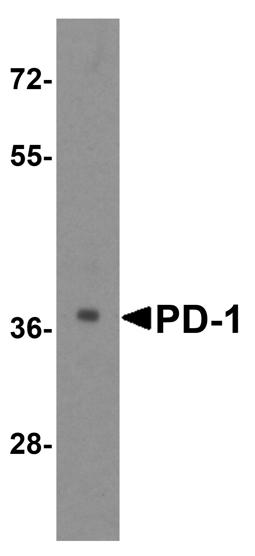
Western blot analysis of PD-1 in transfected 293 cell lysate with PD-1 antibody at 1 µg/mL.
Immunohistochemistry of PD-1 in (A) human tonsil tissue, (B) human lymph node tissue, and (C) human spleen tissue with PD-1 antibody at 5 µg/mL. (D) Immunohistochemistry in human tonsil tissue with control mouse IgG staining at 5 µg/mL.
Immunohistochemistry of PD-1 in (A) human breast cancer tissue and (B) human normal breast tissue with PD-1 antibody at 5 µg/mL.
Immunocytochemistry of PD-1 in transfected 293 cells with PD-1 antibody at 5 µg/mL. Lower left: Immunocytochemistry in transfected 293 cells with control mouse IgG antibody at 5 µg/mL.
Immunofluorescence of PD-1 in transfected 293 cells with PD-1 antibody at 5 µg/mL. Lower left: Immunofluorescence in transfected 293 cells with control mouse IgG antibody at 5 µg/mL. Red: PD1 Antibody [7H6] (orb1239763) Blue: DAPI staining
Western blot analysis of PD-1 in overexpressing 293 cells using orb1239745, orb1239769, and orb1239763 antibody at 1, 0.5, and 0.25 µg/ml, respectively.
Flow cytometry analysis of PD-1 overexpressing 293 cells using orb1239763 at 1 µg/ml. Blue: untransfected cells, Yellow: PD-1 transfected cells.
Titration curve analysis of PD-1 mAbs to detect recombinant PD-1 in ELISA with orb1239745, orb1239769, orb1239763, orb1239738, and orb1239734 abs at decreasing concentrations.
* VAT and and shipping costs not included. Errors and price changes excepted