Anti-PD-1 antibody (orb1239732) was raised against a peptide corresponding to 16 amino acids near the carboxy terminus of human PD-1. The immunogen is located within amino acids 210-260 of PD-1.
Conjugation:
Unconjugated
Alternative Names:
PD-1 Antibody: PD1, PD-1, CD279, SLEB2, hPD-1, hPD-l, hSLE1, PD1, Programmed cell death protein 1, Protein PD-1, PDCD1, PDCD-1
PDCD1 Antibody
Clonality:
Polyclonal
Concentration:
1 mg/mL
Molecular Weight:
Human PD-1 has 1 isoform (288aa, 32kD). Mouse PD-1 has 1 isoform 288aa, 32kD) and Rat PD-1 also has one isoform (287aa, 32kD).
PD-1 Antibody is supplied in PBS containing 0.02% sodium azide.
Form:
Liquid
Target:
PDCD1
KO Validation in HeLa Cells. Loading: 10 µg of HeLa WT cell lysates or PD-1 KO cell lysates. Antibodies: PD-1, orb1239732 (4 µg/mL) and beta-actin orb1240312 (1 µg/mL), 1 h incubation at RT in 5% NFDM/TBST. Secondary: Goat Anti-Rabbit IgG HRP conjugate at 1:10000 dilution.
KD Validation in HeLa Cells. Loading: 10 µg of HeLa WT cell lysates or PD-1 KD cell lysates. Antibodies: PD-1, orb1239732 (4 µg/mL) and beta-actin orb1240312 (1 µg/mL), 1 h incubation at RT in 5% NFDM/TBST. Secondary: Goat Anti-Rabbit IgG HRP conjugate at 1:10000 dilution.
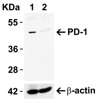
Western Blot Validation in Human and Mouse Cell Lines. Loading: 15 µg of lysates per lane. Antibodies: PD-1 orb1239732 (4 µg/mL), 1h incubation at RT in 5% NFDM/TBST. Secondary: Goat anti-rabbit IgG HRP conjugate at 1:10000 dilution.
Western Blot Validation in THP-1 Cell Lysate in the (A) absence and (B) presence of blocking peptide. Loading: 15 µg of lysates per lane. Antibodies: PD-1, orb1239732 (1 µg/mL), 1h incubation at RT in 5% NFDM/TBST. Secondary: Goat anti-rabbit IgG HRP conjugate at 1:10000 dilution.
Western Blot Validation in Rat Thymus Cell Lysate. Loading: 15 µg of lysates per lane. Antibodies: PD-1 orb1239732 (1 µg/mL), 1h incubation at RT in 5% NFDM/TBST. Secondary: Goat anti-rabbit IgG HRP conjugate at 1:10000 dilution.
Immunohistochemistry Validation of PD-1 in Human Tonsil Tissue. Immunohistochemical analysis of paraffin-embedded human tonsil tissue using anti-PD-1 antibody (orb1239732) at 5 µg/ml. Tissue was fixed with formaldehyde and blocked with 10% serum for 1 h at RT, antigen retrieval was by heat mediation with a citrate buffer (pH6). Samples were incubated with primary antibody overnight at 4C. A goat anti-rabbit IgG H&L (HRP) at 1/250 was used as secondary. Counter stained with Hematoxylin.
Immunohistochemistry Validation of PD-1 in Human Tonsil Tissue. Immunohistochemical analysis of paraffin-embedded human tonsil tissue using anti-PD-1 antibody (orb1239732) at 5 µg/ml. Tissue was fixed with formaldehyde and blocked with 10% serum for 1 h at RT, antigen retrieval was by heat mediation with a citrate buffer (pH6). Samples were incubated with primary antibody overnight at 4C. A goat anti-rabbit IgG H&L (HRP) at 1/250 was used as secondary. Counter stained with Hematoxylin.
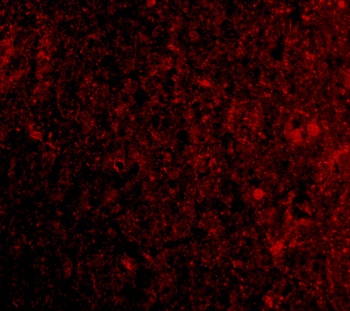
Immunohistochemistry Validation of PD-1 in Human Brain Tissue. Immunohistochemical analysis of paraffin-embedded human brain tissue using anti-PD-1 antibody (orb1239732) at 2.5 µg/ml. Tissue was fixed with formaldehyde and blocked with 10% serum for 1 h at RT, antigen retrieval was by heat mediation with a citrate buffer (pH6). Samples were incubated with primary antibody overnight at 4C. A goat anti-rabbit IgG H&L (HRP) at 1/250 was used as secondary. Counter stained with Hematoxylin.
* VAT and and shipping costs not included. Errors and price changes excepted