Anti-MANF antibody (orb1239620) was raised against a peptide corresponding to 12 amino acids near the amino terminus of human MANF. The immunogen is located within the first 50 amino acids of MANF.
MANF Antibody is supplied in PBS containing 0.02% sodium azide.
Form:
Liquid
Target:
MANF
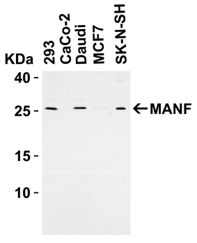
Western Blot Validation in Human Cell Lines. Loading: 15 µg of lysates per lane. Antibodies: MANF orb1239620, (2 µg/mL), 1h incubation at RT in 5% NFDM/TBST. Secondary: Goat anti-rabbit IgG HRP conjugate at 1:10000 dilution.
Western Blot Validation with Recombinant Protein. Loading: 30 ng of human MANF recombinant protein per lane. Antibodies: MANF orb1239620 (Lane 1: 0.125 µg/mL, Lane 2: 0.25 µg/mL and Lane 3: 0.5 µg/mL), 1h incubation at RT in 5% NFDM/TBST. Secondary: Goat anti-rabbit IgG HRP conjugate at 1:10000 dilution. Observed at around 55kD.
Western Blot Validation in Rat Brain Tissue Lysate. Loading: 15 µg of lysates per lane. Antibodies: MANF orb1239620 (A: 1 µg /mL and B: 2 µg /mL), 1h incubation at RT in 5% NFDM/TBST. Secondary: Goat anti-rabbit IgG HRP conjugate at 1:10000 dilution.
Immunohistochemistry Validation of MANF in Human Brain Tissue. Immunohistochemical analysis of paraffin-embedded human brain tissue using anti-MANF antibody (orb1239620) at 2.5 µg /ml. Tissue was fixed with formaldehyde and blocked with 10% serum for 1 h at RT, antigen retrieval was by heat mediation with a citrate buffer (pH6). Samples were incubated with primary antibody overnight at 4C. A goat anti-rabbit IgG H&L (HRP) at 1/250 was used as secondary. Counter stained with Hematoxylin.
Immunofluorescence Validation of MANF in Human Brain Tissue. Immunofluorescent analysis of 4% paraformaldehyde-fixed human brain tissue labeling MANF with orb1239620 at 20 µg /mL, followed by goat anti-rabbit IgG secondary antibody at 1/500 dilution (red).
Induced Expression Validation of MANF in Mouse C17.2 Neural Stem Cells (Almutawaa et al., 2014). Concentration- and time-dependent effects of Valproic acid (VPA) on MANF at 48 h examined by Western blot analysis with anti-MANF antibodies. MANF was markedly increased 48h after VPA treatment. Lanes 1-4: control, 0.5 mM VPA, 1 mM VPA, 3 mM VPA.
* VAT and and shipping costs not included. Errors and price changes excepted