CIDE-A antibody was raised against an 18 amino acid peptide near the carboxy terminus of mouse CIDE-A.The immunogen is located within the last 50 amino acids of CIDE-A.
Conjugation:
Unconjugated
Alternative Names:
CIDE-A Antibody: CIDE-A, Cell death activator CIDE-A, Cell death-inducing DFFA-like effector A
CIDE-A Antibody is supplied in PBS containing 0.02% sodium azide.
Form:
Liquid
Target:
CIDEA
Application Notes:
Application Notes: CIDE-A antibody can be used for detection of CIDE-A by Western blot at 0.5 µg/mL. Antibody can also be used for immunohistochemistry starting at 2.5 µg/mL. For immunofluorescence start at 20 µg/mL.Antibody validated: Western Blot in mouse samples, Immunohistochemistry in human and mouse samples and Immunofluorescence in human and mouse samples. All other applications and species not yet tested
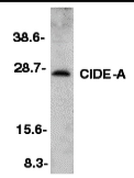
Western blot analysis of CIDE-A in mouse heart tissue lysate with CIDE-A antibody at 1:500 dilution.
Immunohistochemistry of CIDE-A in mouse heart tissue with CIDE-A antibody at 5 µg/mL.
Immunofluorescence of CIDE-A in Mouse Heart cells with CIDE-A antibody at 20 ug/mL.
Immunohistochemistry of CIDE-A in human brain tissue with CIDE-A antibody at 2.5 µg/ml.
Immunofluorescence of CIDE-A in human brain tissue with CIDE A antibody at 20 µg/mL. Green: CIDE-A Antibody (orb1239296) Blue: DAPI staining.
Immunohistochemistry of CIDE-A in human brain tissue with CIDE A antibody at 2.5 µg/mL.
* VAT and and shipping costs not included. Errors and price changes excepted