E.coli-derived human Daxx recombinant protein (Position: K56-R345). Human Daxx shares 88.3% and 88.6% amino acid (aa) sequence identity with mouse and rat Daxx, respectively.
Alternative Names:
BING2, DAP6, DAXX, EAP1, ETS1 associated protein 1, hDaxx
Flow Cytometry, Optimal dilutions should be determined by end users.
IHC analysis of DAXX using anti-DAXX antibody (PB9550). DAXX was detected in a paraffin-embedded section of rat intestine tissue. Heat mediated antigen retrieval was performed in EDTA buffer (pH 8.0, epitope retrieval solution). The tissue section was blocked with 10% goat serum. The tissue section was then incubated with 1 µg/ml rabbit anti-DAXX Antibody (PB9550) overnight at 4C. Biotinylated goat anti-rabbit IgG was used as secondary antibody and incubated for 30 minutes at 37C. The tissue section was developed using Strepavidin-Biotin-Complex (SABC) (Catalog SA1022) with DAB as the chromogen.
IHC analysis of DAXX using anti-DAXX antibody (PB9550). DAXX was detected in a paraffin-embedded section of human intestinal cancer tissue. Heat mediated antigen retrieval was performed in EDTA buffer (pH 8.0, epitope retrieval solution). The tissue section was blocked with 10% goat serum. The tissue section was then incubated with 1 µg/ml rabbit anti-DAXX Antibody (PB9550) overnight at 4C. Biotinylated goat anti-rabbit IgG was used as secondary antibody and incubated for 30 minutes at 37C. The tissue section was developed using Strepavidin-Biotin-Complex (SABC) (Catalog SA1022) with DAB as the chromogen.
IHC analysis of DAXX using anti-DAXX antibody (PB9550). DAXX was detected in immunocytochemical section of SMMC-7721 Cell. Enzyme antigen retrieval was performed using IHC enzyme antigen retrieval reagent (AR0022) for 15 mins. The cells were blocked with 10% goat serum. And then incubated with 1µg/ml rabbit anti-DAXX Antibody (PB9550) overnight at 4C. Biotinylated goat anti-rabbit IgG was used as secondary antibody and incubated for 30 minutes at 37C. The section was developed using Strepavidin-Biotin-Complex (SABC)(Catalog SA1022) with DAB as the chromogen.
IHC analysis of DAXX using anti-DAXX antibody (PB9550). DAXX was detected in immunocytochemical section of A549 Cell. Enzyme antigen retrieval was performed using IHC enzyme antigen retrieval reagent (AR0022) for 15 mins. The cells were blocked with 10% goat serum. And then incubated with 1µg/ml rabbit anti-DAXX Antibody (PB9550) overnight at 4C. Biotinylated goat anti-rabbit IgG was used as secondary antibody and incubated for 30 minutes at 37C. The section was developed using Strepavidin-Biotin-Complex (SABC)(Catalog SA1022) with DAB as the chromogen.
IF analysis of Daxx using anti-Daxx antibody (PB9550). Daxx was detected in immunocytochemical section of U20S cells. Enzyme antigen retrieval was performed using IHC enzyme antigen retrieval reagent (AR0022) for 15 mins. The cells were blocked with 10% goat serum. And then incubated with 2µg/mL rabbit anti-Daxx Antibody (PB9550) overnight at 4C. DyLight488 Conjugated Goat Anti-Rabbit IgG (BA1127) was used as secondary antibody at 1:100 dilution and incubated for 30 minutes at 37C. T
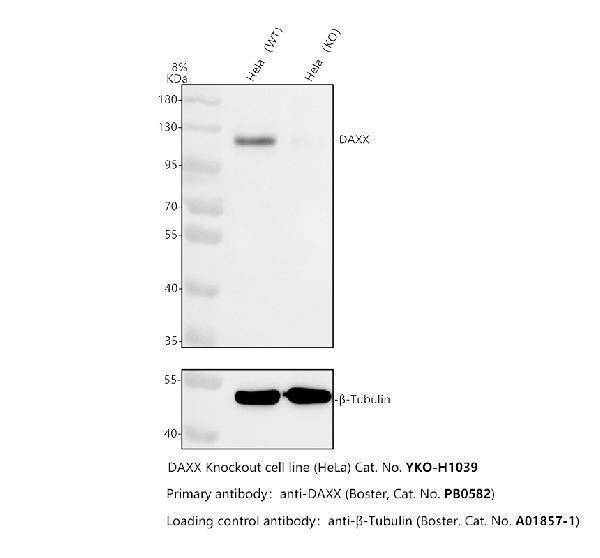
Western blot analysis of Daxx using anti-Daxx antibody (PB9550). Electrophoresis was performed on a 5-20% SDS-PAGE gel at 70V (Stacking gel) / 90V (Resolving gel) for 2-3 hours. The sample well of each lane was loaded with 30 ug of sample under reducing conditions. Lane 1: human K562 whole cell lysates, Lane 2: rat PC-12 whole cell lysates, Lane 3: mouse thymus tissue lysates. After electrophoresis, proteins were transferred to a nitrocellulose membrane at 150 mA for 50-90 minutes. Blocked the membrane with 5% non-fat milk/TBS for 1.5 hour at RT. The membrane was incubated with rabbit anti-Daxx antigen affinity purified polyclonal antibody (Catalog PB9550) at 0.5 µg/mL overnight at 4C, then washed with TBS-0.1%Tween 3 times with 5 minutes each and probed with a goat anti-rabbit IgG-HRP secondary antibody at a dilution of 1:5000 for 1.5 hour at RT. The signal is developed using an Enhanced Chemiluminescent detection (ECL) kit (Catalog EK1002) with Tanon 5200 system. A specific band was detected for Daxx at approximately 115 kDa. The expected band size for Daxx is at 81 kDa.
* VAT and and shipping costs not included. Errors and price changes excepted