Rabbit IgG in stabilizing components, phosphate buffered saline, pH 7.4, 150mM NaCl, 0.02% sodium azide and 50% glycerol. *This antibody is supplied in a stabilized formulation. Compatibility with conjugation reactions depends on the chemistry of the con
Purity:
Affinity-chromatography
Form:
Liquid
Target:
Growth/differentiation factor 15
Application Dilute:
WB 1:500-2000
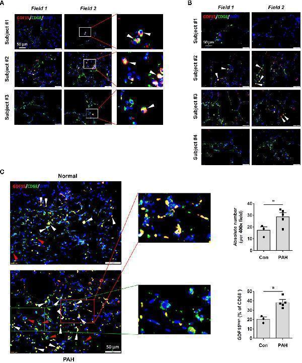
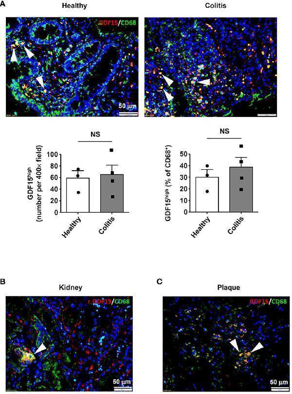
Immunofluorescence double labeling showing the existence of GDF15 high macrophages in lung tissues. (A) Results obtained in healthy human lung tissues from 3 independent subjects. Two representative microscopic fields were shown. Arrowheads indicated the CD68 + GDF15 high macrophages. (B) Results obtained in human lung tissues with COPD from 4 independent subjects. Arrowheads indicated the CD68 + GDF15 high macrophages. (C) Results obtained in rat lung tissues without and with experimental PAH. The nuclei were counterstained with DAPI (blue). White arrowheads indicated CD68 + GDF15 high macrophages. The red box highlighted the presence of CD68 + GDF15 high macrophages (white arrowheads), the green box highlighted the presence of CD68 + GDF15 low macrophages (red arrowheads). The bar graphs showed the absolute and relative abundances of CD68 + GDF15 high macrophages in normal and PAH lungs. Data were expressed as mean SEM. * P < 0.05, unpaired t -test.Index in PubMed under a CC BY license. PMID: 38655264
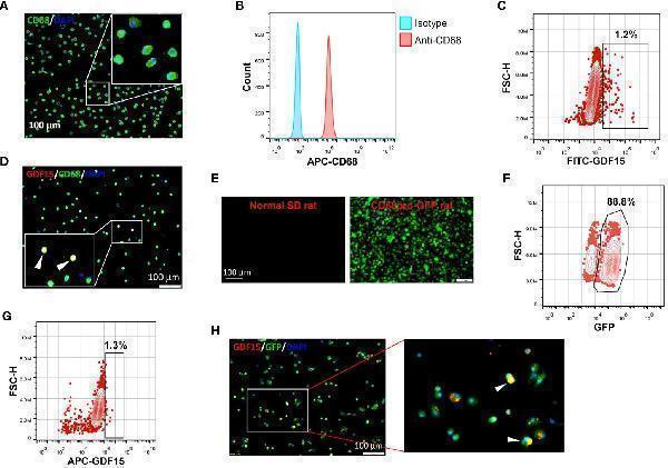
GDF15 high macrophages could be derived by in vitro differentiation of mononuclear cells. (A, B) Immunofluorescence staining and flow cytometry results confirming that in vitro differentiation of human peripheral blood mononuclear cells (PBMNCs) with GM-CSF for 7 days yielded CD68 + macrophages. (C, D) Flow cytometry and immunofluorescence double labeling results showing that the PBMNC-derived macrophages contained a minor population of GDF15 high cells (arrowheads in D ) (example from 3 independent experiments). (E, F) Fluorescence microscopy and flow cytometry data showing that GM-CSF differentiation of rat bone marrow mononuclear cells (BMMNCs) in vitro yielded macrophages (GFP expressing) of a high purity (~90%). CD68pro-GFP rats had a GFP transgene under the control of CD68 promoter. Cells from normal rats showing no GFP fluorescence served as a negative control (left panel in E ). (G, H) Flow cytometry and immunofluorescence double labeling data (from 3 independent experiments) showing that the BMMNC-derived macrophages (from CD68pro-GFP rats) contained a minor population of GDF15 high cells (arrowheads in H ). The flow cytometry data in panels (C, G) were from cells gated for GFP + . The nuclei were counterstained with DAPI (blue).Index in PubMed under a CC BY license. PMID: 38655264
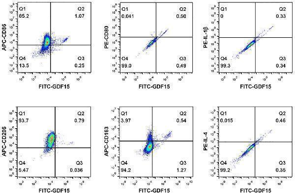
Flow cytometry results showing that GDF15 high macrophages did not exhibit a typical M1 or M2 phenotype. Experiments were performed in human PBMNC-derived macrophages, using CD86, CD80 and IL-1beta as the M1 markers, and CD206, CD163 and IL-4 as the M2 markers. Data were from a single test using pooled samples from 4 healthy volunteers.Index in PubMed under a CC BY license. PMID: 38655264
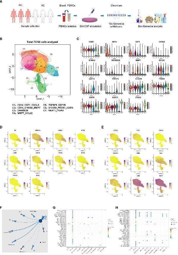
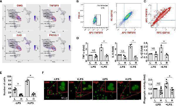
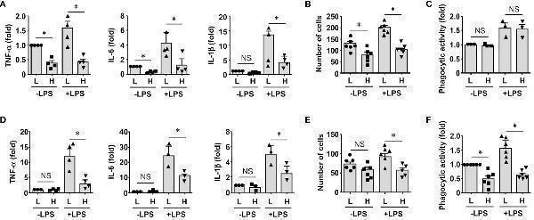
Molecular characterization of human PBMNC-derived GDF15 high macrophages with scRNA-seq. (A) Graphical outline of the experimental procedure. (B) UMAP plots showing the identified cell sub-populations (C1 to C7) based on the scRNA-seq data from total 73,768 cells pooled from samples of 3 healthy volunteers, 3 PAH patients harboring mutations in BMPR2 gene, and 3 PAH patients without BMPR2 mutations. The putative nomenclatures for C1 to C7 were given below the graph. The numbers 0 to 13 demarcated the initial cell clusters obtained with the default clustering process of Seurat. (C) Violin plots showing expression patterns of the identified marker genes for C1 to C7. The horizontal bars represented median values. (D) UMAP plots showing expression patterns of the top 10 genes that were overexpressed in GDF15 high macrophages (C5) as compared to GDF15 low c
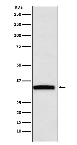
Western blot analysis of GDF15 expression in HepG2 cell lysate.
* VAT and and shipping costs not included. Errors and price changes excepted