A synthesized peptide derived from human Met (c-Met)
Alternative Names:
AUTS9, c-Met, c-Met (Cytoplasmic), HGF receptor, HGF/SF receptor, HGFR, MET, Proto oncogene c Met, RCCP2, Scatter factor receptor, SF receptor, Tyrosine protein kinase Met
Boster Bio Anti-Met (c-Met) Rabbit Monoclonal Antibody catalog M01488. Tested in WB, IHC, ICC/IF, Flow Cytometry applications. This antibody reacts with Human, Mouse, Rat.
Rabbit IgG in stabilizing components, phosphate buffered saline, pH 7.4, 150mM NaCl, 0.02% sodium azide and 50% glycerol. *This antibody is supplied in a stabilized formulation. Compatibility with conjugation reactions depends on the chemistry of the con
Purity:
Affinity-chromatography
Form:
Liquid
Target:
Hepatocyte growth factor receptor
Application Dilute:
WB 1:500-2000IHC 1:50-200ICC/IF 1:50-200FC 1:50
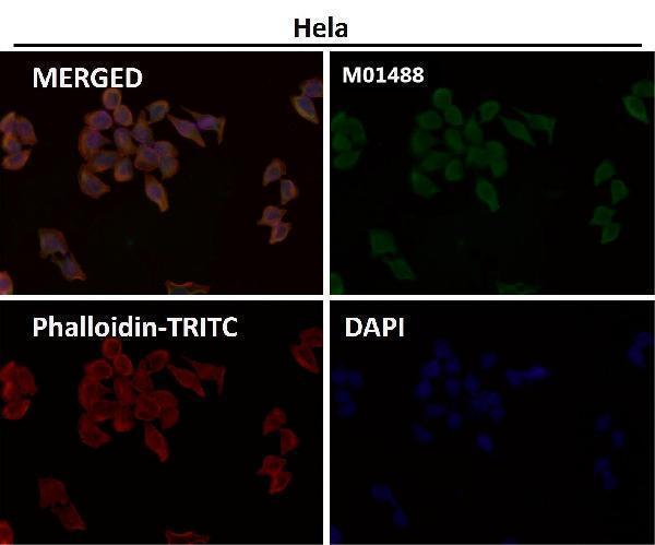
Immunofluorescent analysis using the Antibody at 1:50 dilution.
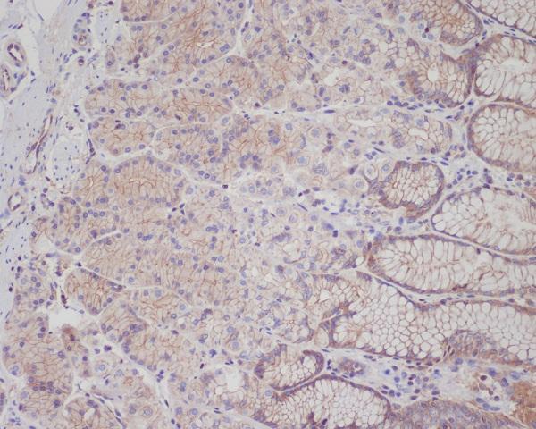
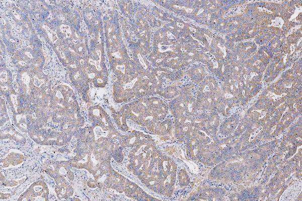
Immunohistochemical analysis of paraffin-embedded human stomach, using Met (c-Met) Antibody .
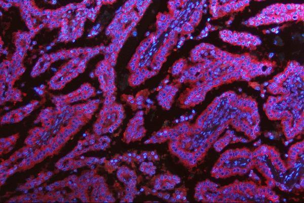
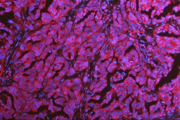
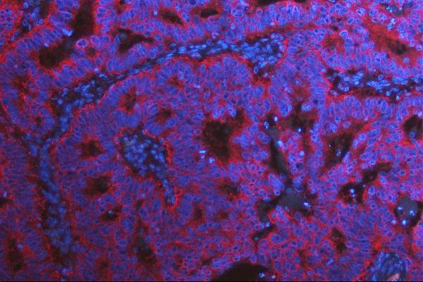
IF analysis of MET using anti-MET antibody (M01488). MET was detected in a paraffin-embedded section of human lung cancer tissue. Heat mediated antigen retrieval was performed in EDTA buffer (pH 8.0, epitope retrieval solution). The tissue section was blocked with 10% goat serum. The tissue section was then incubated with 5 µg/mL rabbit anti-MET Antibody (M01488) overnight at 4C. DyLight594 Conjugated Goat Anti-Rabbit IgG (BA1142) was used as secondary antibody at 1:100 dilution and incubated for 30 minutes at 37C. The section was counterstained with DAPI. Visualize using a fluorescence microscope and filter sets appropriate for the label used.
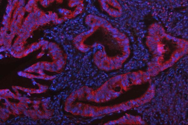
IF analysis of MET using anti-MET antibody (M01488). MET was detected in a paraffin-embedded section of human colon cancer tissue. Heat mediated antigen retrieval was performed in EDTA buffer (pH 8.0, epitope retrieval solution). The tissue section was blocked with 10% goat serum. The tissue section was then incubated with 5 µg/mL rabbit anti-MET Antibody (M01488) overnight at 4C. DyLight594 Conjugated Goat Anti-Rabbit IgG (BA1142) was used as secondary antibody at 1:100 dilution and incubated for 30 minutes at 37C. The section was counterstained with DAPI. Visualize using a fluorescence microscope and filter sets appropriate for the label used.
IF analysis of MET using anti-MET antibody (M01488). MET was detected in a paraffin-embedded section of human ovarian cancer tissue. Heat mediated antigen retrieval was performed in EDTA buffer (pH 8.0, epitope retrieval solution). The tissue section was blocked with 10% goat serum. The tissue section was then incubated with 5 µg/mL rabbit anti-MET Antibody (M01488) overnight at 4C. DyLight594 Conjugated Goat Anti-Rabbit IgG (BA1142) was used as secondary antibody at 1:100 dilution and incubated for 30 minutes at 37C. The section was counterstained with DAPI. Visualize using a fluorescence microscope and filter sets appropriate for the label used.
IF analysis of MET using anti-MET antibody (M01488). MET was detected in a paraffin-embedded section of human stomach cancer tissue. Heat mediated antigen retrieval was performed in EDTA buffer (pH 8.0, epitope retrieval solution). The tissue section was blocked with 10% goat serum. The tissue section was then incubated with 5 µg/mL rabbit anti-MET Antibody (M01488) overnight at 4C. DyLight594 Conjugated Goat Anti-Rabbit IgG (BA1142) was used as secondary antibody at 1:100 dilution and incubated for 30 minutes at 37C. The section was counterstained with DAPI. Visualize using a fluorescence microscope and filter sets appropriate for the label used.
IHC analysis of MET using anti-MET antibody (M01488). MET was detected in a paraffin-embedded section of human stomach cancer tissue. Heat mediated antigen retrieval was performed in EDTA buffer (pH 8.0, epitope retrieval solution). The tissue section was blocked with 10% goat serum. The tissue section was then incubated with 5 µg/ml rabbit anti-MET Antibody (M01488) overnight at 4C. HRP Conjugated Goat Anti-rabbit IgG was used as secondary antibody and incubated for 30 minutes at 37C. The tissue section was developed using HRP Conjugated Rabbit IgG Super Vision Assay Kit (Catalog SV0002) with DAB as the chromogen.
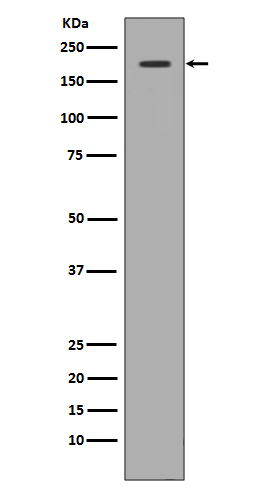
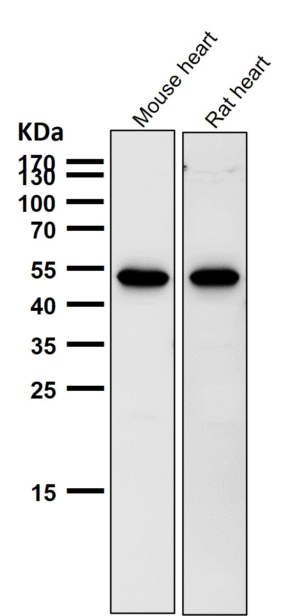
Western blot analysis of c-Met expression in 293 cell lysate.
All lanes use the Antibody at 1:1K dilution for 1 hour at room temperature.
* VAT and and shipping costs not included. Errors and price changes excepted