PBS (pH 7.3) containing 1% stabilizing protein, 50% glycerol and 0.02% sodium azide.This antibody is supplied in a stabilized formulation. Compatibility with conjugation reactions depends on the chemistry of the conjugation method used. For conjugation me
Application Dilute:
WB 1:2000IHC 1:150IF 1:100Flow Cytometry 1:100
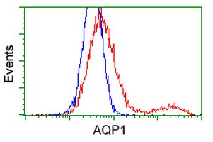
HEK293T cells transfected with either AQP1 (Myc-DDK-tagged) overexpress plasmid (Red) or empty vector control plasmid (Blue) were immunostained by anti-AQP1 antibody (M00865-1)
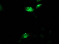
Anti-AQP1 mouse monoclonal antibody (M00865-1) immunofluorescent staining of COS7 cells transiently transfected by pCMV6-ENTRY AQP1.
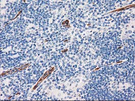
Immunohistochemical staining of paraffin-embedded Human lymphoma tissue using anti-AQP1 mouse monoclonal antibody. (Heat-induced epitope retrieval by 10mM citric buffer
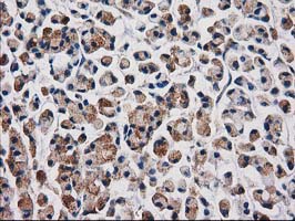
Immunohistochemical staining of paraffin-embedded Adenocarcinoma of Human colon tissue using anti-AQP1 mouse monoclonal antibody. (Heat-induced epitope retrieval by 10mM citric buffer
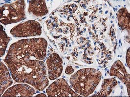
Immunohistochemical staining of paraffin-embedded Human Kidney tissue within the normal limits using anti-AQP1 mouse monoclonal antibody. (Heat-induced epitope retrieval by 10mM citric buffer
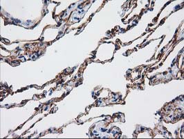
Immunohistochemical staining of paraffin-embedded Human lung tissue within the normal limits using anti-AQP1 mouse monoclonal antibody. (Heat-induced epitope retrieval by 10mM citric buffer
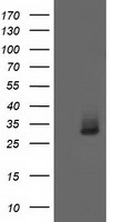
HEK293T cells were transfected with the pCMV6-ENTRY control (Left lane) or pCMV6-ENTRY AQP1 (Right lane) cDNA for 48 hrs and lysed. Equivalent amounts of cell lysates (5 ug per lane) were separated by SDS-PAGE and immunoblotted with anti-AQP1.
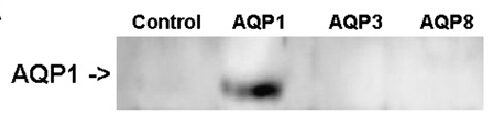
Figure from citation: Western Blot of AQP1 protein level by using anti-AQP1 antibody in human B1647 cells.
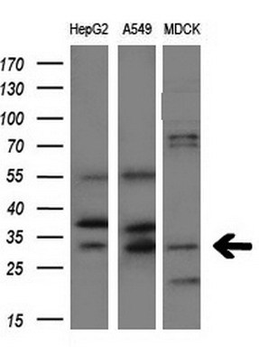
Western blot analysis of extracts (10ug) from 3 different cell lines by using anti-AQP1 monoclonal antibody at 1:200 dilution.
* VAT and and shipping costs not included. Errors and price changes excepted