Boster Bio Anti-LRPPRC/GP130 Antibody Picoband catalog A03264. Tested in ELISA, Flow Cytometry, IF, IHC, ICC, WB applications. This antibody reacts with Human, Monkey, Mouse, Rat. The brand Picoband indicates this is a premium antibody that guarantees superior quality, high affinity, and strong signals with minimal background in Western blot applications. Only our best-performing antibodies are designated as Picoband, ensuring unmatched performance.
Clonality:
Polyclonal
Concentration:
Adding 0.2 ml of distilled water will yield a concentration of 500 µg/ml.
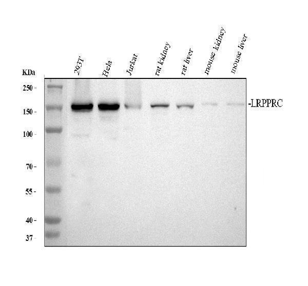
Western blot, 0.1-0.25µg/ml, Human, Mouse, Rat, Monkey Immunohistochemistry (Paraffin-embedded Section), 0.5-1µg/ml, Human, Mouse, Rat Immunocytochemistry/Immunofluorescence, 2µg/ml, Human Flow Cytometry (Fixed), 1-3µg/1x106 cells, Human, Rat ELISA, 0.1-0
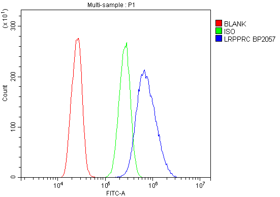
Flow Cytometry analysis of A431 cells using anti-LRPPRC antibody (A03264). Overlay histogram showing A431 cells stained with A03264 (Blue line). To facilitate intracellular staining, cells were fixed with 4% paraformaldehyde and permeabilized with permeabilization buffer. The cells were blocked with 10% normal goat serum. And then incubated with rabbit anti-LRPPRC Antibody (A03264, 1µg/1x106 cells) for 30 min at 20C. DyLight488 conjugated goat anti-rabbit IgG (BA1127, 5-10µg/1x106 cells) was used as secondary antibody for 30 minutes at 20C. Isotype control antibody (Green line) was rabbit IgG (1µg/1x106) used under the same conditions. Unlabelled sample without incubation with primary antibody and secondary antibody (Red line) was used as a blank control.
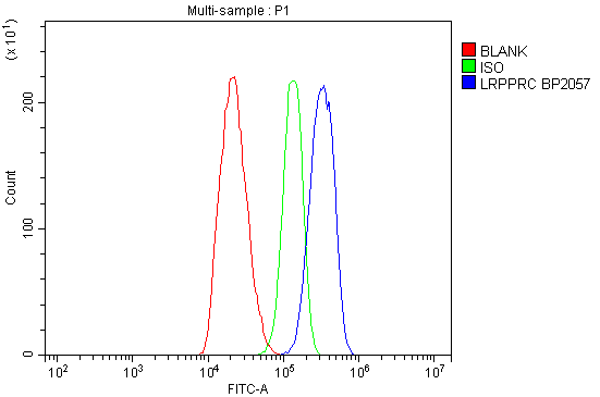
Flow Cytometry analysis of C6 cells using anti-LRPPRC antibody (A03264). Overlay histogram showing C6 cells stained with A03264 (Blue line). To facilitate intracellular staining, cells were fixed with 4% paraformaldehyde and permeabilized with permeabilization buffer. The cells were blocked with 10% normal goat serum. And then incubated with rabbit anti-LRPPRC Antibody (A03264, 1µg/1x106 cells) for 30 min at 20C. DyLight488 conjugated goat anti-rabbit IgG (BA1127, 5-10µg/1x106 cells) was used as secondary antibody for 30 minutes at 20C. Isotype control antibody (Green line) was rabbit IgG (1µg/1x106) used under the same conditions. Unlabelled s
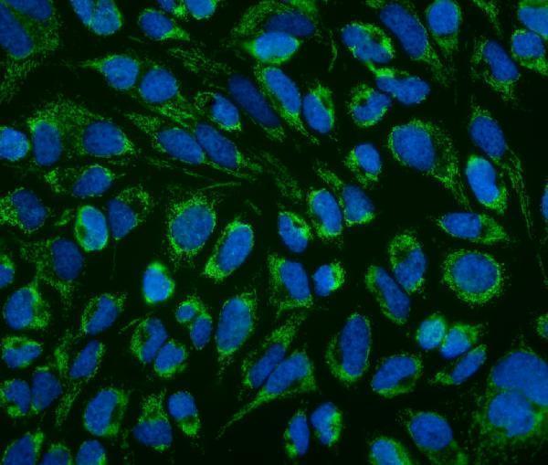
IF analysis of LRPPRC using anti-LRPPRC antibody (A03264). LRPPRC was detected in immunocytochemical section of Hela cells. Enzyme antigen retrieval was performed using IHC enzyme antigen retrieval reagent (AR0022) for 15 mins. The cells were blocked with 10% goat serum. And then incubated with 2µg/mL rabbit anti-LRPPRC Antibody (A03264) overnight at 4C. DyLight488 Conjugated Goat Anti-Rabbit IgG (BA1127) was used as secondary antibody at 1:100 dilution and incubated for 30 minutes at 37C. The section was counterstained with DAPI. Visualize using a fluorescence microscope and filter sets appropriate for the label used.
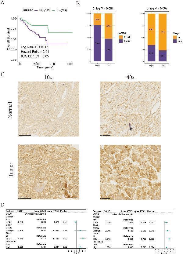
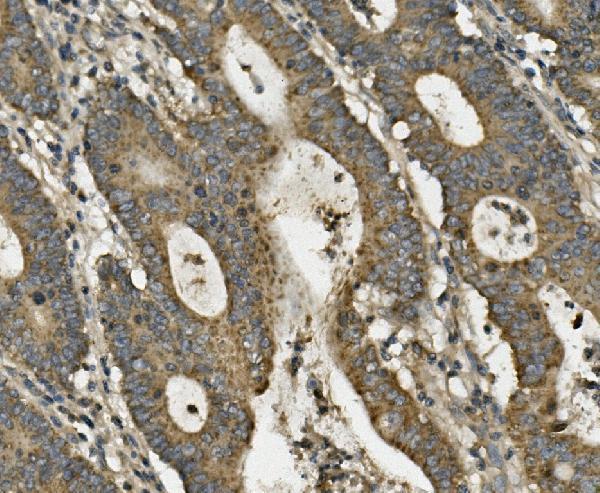
IHC analysis of LRPPRC using anti-LRPPRC antibody (A03264). LRPPRC was detected in paraffin-embedded section of human rectal cancer tissue. Heat mediated antigen retrieval was performed in EDTA buffer (pH8.0, epitope retrieval solution). The tissue section was blocked with 10% goat serum. The tissue section was then incubated with 1µg/ml rabbit anti-LRPPRC Antibody (A03264) overnight at 4C. Biotinylated goat anti-rabbit IgG was used as secondary antibody and incubated for 30 minutes at 37C. The tissue section was developed using Strepavidin-Biotin-Complex (SABC) (Catalog SA1022) with DAB as the chromogen.
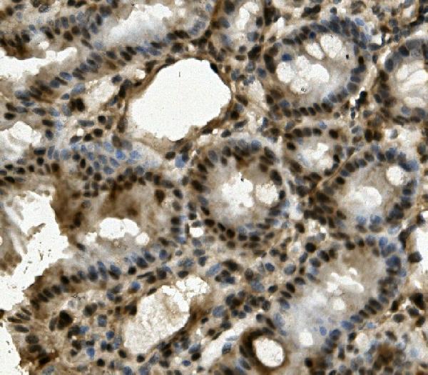
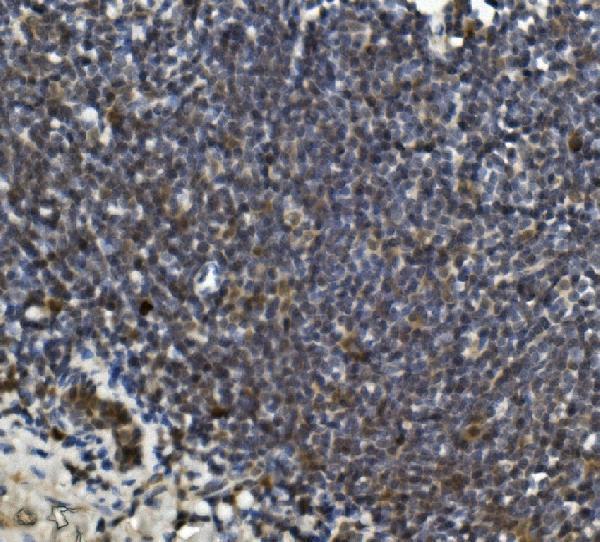
IHC analysis of LRPPRC using anti-LRPPRC antibody (A03264). LRPPRC was detected in paraffin-embedded section of mouse intestine tissue. Heat mediated antigen retrieval was performed in EDTA buffer (pH8.0, epitope retrieval solution). The tissue section was blocked with 10% goat serum. The tissue section was then incubated with 1µg/ml rabbit anti-LRPPRC Antibody (A03264) overnight at 4C. Biotinylated goat anti-rabbit IgG was used as secondary antibody and incubated for 30 minutes at 37C. The tissue section was developed using Strepavidin-Biotin-Complex (SABC) (Catalog SA1022) with DAB as the chromogen.
IHC analysis of LRPPRC using anti-LRPPRC antibody (A03264). LRPPRC was detected in paraffin-embedded section of rat intestine tissue. Heat mediated antigen retrieval was performed in EDTA buffer (pH8.0, epitope retrieval solution). The tissue section was blocked with 10% goat serum. The tissue section was then incubated with 1µg/ml rabbit anti-LRPPRC Antibody (A03264) overnight at 4C. Biotinylated goat anti-rabbit IgG was used as secondary antibody and incubated for 30 minutes at 37C. The tissue section was developed using Strepavidin-Biotin-Complex (SABC) (Catalog SA1022) with DAB as the chromogen.
* VAT and and shipping costs not included. Errors and price changes excepted