
Monoclonal Antibody to FSH Receptor (Clone: ABM5D29)
Catalog Number:
ABI-10-7600-25
- Images (3)

| Article Name: | Monoclonal Antibody to FSH Receptor (Clone: ABM5D29) |
| Biozol Catalog Number: | ABI-10-7600-25 |
| Supplier Catalog Number: | 10-7600-25 |
| Alternative Catalog Number: | ABI-10-7600-25-25UG |
| Manufacturer: | Abeomics |
| Host: | Mouse |
| Category: | Antikörper |
| Application: | FACS, WB |
| Species Reactivity: | Human |
| Immunogen: | A partial length recombinant protein (a.a 03-200) of FSH Receptor was used as the immunogen for this antibody. |
| Alternative Names: | Follicle-stimulating hormone receptor, FSH-R, Follitropin receptor |
| FSH receptor is a member of the G protein coupled receptor family consisting of 9 introns and 10 exons and a promoter region and is located on chromosome 2p21. FSH receptor was shown to be expressed selectively on the luminal surface of tumor blood vessels. A general characteristic of the blood vessels that express the endothelial FSH receptor is that they are located at the periphery of the tumors. Many studies showed that FSH receptor polymorphisms at positions 307 and 680 may be even more relevant for clinical practice for the reasons of regulating the ovarian reaction to hormone, controlling ovarian hyperstimulation, changing the menstrual cycle, and causing POF (Premature Ovarian Failure) and PCOS (Polycystic Ovary Syndrome). |
| Application Dilute: | WB: 2-4 µg/ml, FACS: 0.5-1 µg/10 6 |
|
|
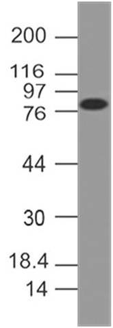
Fig-1: Western blot analysis of FSH Receptor. Anti-FSH Receptor antibody (Clone: ABM5D29) was used at 2 µg/ml on Hela lysate. |
|
|
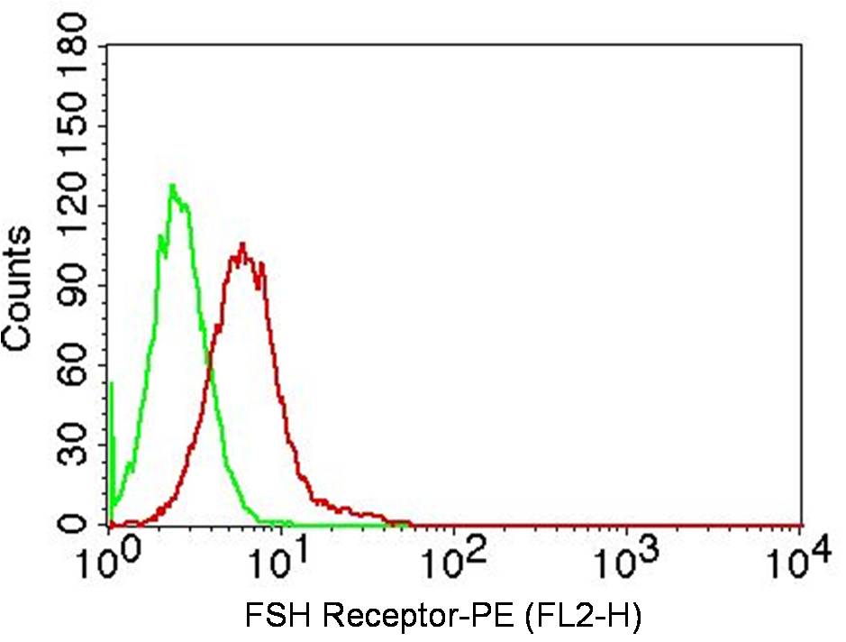
Fig-2: Intracellular flow analysis of FSH receptor antibody on A431 cells using 0.5 µg/10 6 cells (Clone: ABM5D29). Green represents isotype control, red represents anti-FSH receptor antibody. Goat anti-mouse PE conjugate was used as secondary antibody. (Cells were fixed with 4% paraformaldehyde for 10 min and washed with PBS by centrifuging at 1100 for 5 min followed by permeabilization for 20 min and washed again as mentioned above. Then cell were incubated with primary antibody for 45 min. |
|
|
Fig-3: Intracellular flow analysis of FSH receptor antibody on HeLa cells using 0.5 µg/10 6 cells (Clone: ABM5D29). Green represents isotype control, red represents anti-FSH receptor antibody. Goat anti-mouse PE conjugate was used as secondary. (Cells were fixed with 4% paraformaldehyde for 10 min and washed with PBS by centrifuging at 1100 for 5 min followed by permeabilization for 20 min and washed again as mentioned above. Then cell were incubated with primary antibody for 45 min. and afte |
Product Guarantee and Expert Support
