The immunogen for this CD56 antibody was a membrane preparation of a small cell lung carcinoma.
Alternative Names:
NCAM1||NCAM
CD56, a 175-220KDa glycoprotein, is a member of the Ig super family. It is expressed as three major isoforms and consists of five Ig-like domains and two Fibronectin-type III domains in the extracellular region. The 135kDa isoform is the basic molecule which is glycosylated or sialylated to produce the mature species. CD56 is widely expressed in nervous system, on NK cells and a specific set of Tcells. CD56+ NK cells and Tcells are unique in their ability to mediate cell-mediated cytotoxicity against certain tumor cell targets without MHC restriction. Other physiological functions of CD56 include mediating cell adhesion through homophilic and heterophilic interaction and activating intracellular signaling pathways resulting in neutrite extension and fasciculation, migration and synapses formation in brain. CD56 is also vital for neuronal development and plasticity in adult brain. It is used as a tumor marker in various cancers such as NK lymphomas and Merkel cell carcinoma. NCAM is expressed on most neuroectodermal derived lines, tissues, and neoplasms such as retinoblastoma, medulloblastoma, astrocytoma, and neuroblastoma. It is also expressed on some mesodermally derived tumors such as rhabdomyosarcoma and also on natural killer cells.
Fig: 3 Immunohistochemical analysis of CD56 in human Brain, Cortex tissue using CD56 antibody (Clone: 123C3) at 10 µg/ml.
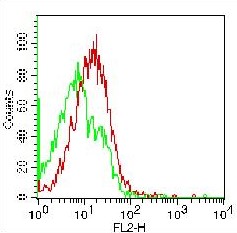
Fig: 1 Flow Cytometric analysis of hCD56 on human PBMC, lymphocytes using 1 µg/10 6 cells. Green represents isotype control (ABEOMICS), red represents anti-hCD56 antibody (10-7504). Goat anti-mouse PE conjugated secondary antibody (ABEOMICS) was used.
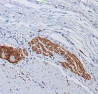
Fig: 2 Immunohistochemical analysis of CD56 in human colon ganglion using CD56 antibody (Clone: 123C3) at 1:200 dilution.
* VAT and and shipping costs not included. Errors and price changes excepted