NALE(TM) Monoclonal antibody to B7-H4 (Clone: ABM53A6) (No Azide, Low Endotoxin)
Biozol Catalog Number:
ABI-10-4172-NALE-25
Supplier Catalog Number:
10-4172-NALE-25
Alternative Catalog Number:
ABI-10-4172-NALE-25-25UG
Manufacturer:
Abeomics
Host:
Mouse
Category:
Antikörper
Application:
FACS, IHC, WB
Species Reactivity:
Human, Mouse
Immunogen:
A partial length recombinant protein (a.a 23-220) of B7-H4 was used as the immunogen for this antibody.
Conjugation:
Unconjugated
Alternative Names:
V-set domain-containing T-cell activation inhibitor 1, B7 homolog 4, B7h.5, Immune costimulatory protein B7-H4, Protein B7S1
B7-H4, a member of B7 family, is a transmembrane protein that has been shown to inhibit T cell responses and neutrophil expansion during bacterial infections. B7-H4 mRNA is widely distributed in mouse and human peripheral tissues and can be induced in monocytes, macrophages, and dendritic cells upon IL-6 and IL-10 stimulation. However, in a variety of tumor cells, B7-H4 is predominantly present in intracellular compartments with unknown mechanism and functions. Cell surface expression of B7-H4 protein is limited and shows an inducible pattern on hematopoietic cells. The overexpression of B7-H4 has been found in various types of human tumors, such as breast cancer, renal cell carcinoma (RCC), ovarian cancer, esophageal squamous cell carcinoma, gastric cancer, pancreatic cancer and melanoma etc, where its expression level is positively correlated with disease progression. By arresting cell cycle, B7-H4 ligation of T cells has a profound inhibitory effect on the growth, cytokine secretion, and development of cytotoxicity.
FACS: 0.5-1 µg/10 6 cells, Immunohistochemical analysis: 5 µg/ml, Western blot analysis: 2-4 µg/ml
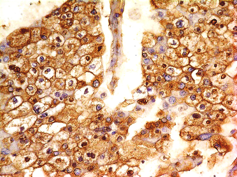
Fig-1: Immunohistochemical analysis of B7-H4 antibody in Renal cell carcinoma using anti-B7-H4 antibody (Clone: ABM53A6) at 5 µg/ml
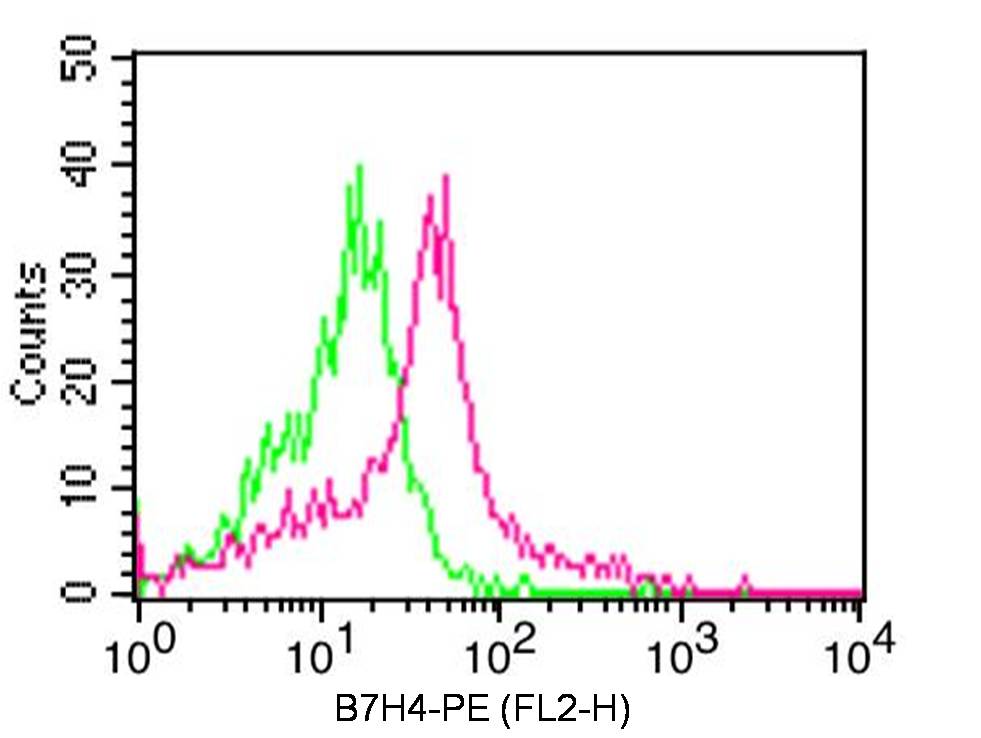
Fig-2: Cell surface Flow analysis of B7-H4 antibody in human PBMC (Monocytes) cells using 0.5 µg/ 10 6 cells of anti-B7-H4 antibody (ABM53A6). Green represents isotype control,red represents anti-B7H4 antibody. Goat anti-mouse PE conjugate was used as secondary antibody.
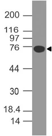
Fig-3: Western blot analysis of B7-H4. Anti-B7-H4 antibody (Clone: ABM53A6) was used at 4 µg/ml on human B7H4-FC fusion protein Lysate.
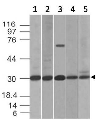
Fig-4: Western blot analysis of B7-H4. Anti-B7-H4 antibody (Clone: ABM53A6) was used at 2 µg/ml on (1) HCT-116, (2) PC3, (3) Kato 111, (4) C2C12 and (5) RAW Lysates.
* VAT and and shipping costs not included. Errors and price changes excepted