
PRP19 Rabbit mAb, Unconjugated, Monoclonal
Catalog Number:
ABB-A9660
- Images (8)

| Article Name: | PRP19 Rabbit mAb, Unconjugated, Monoclonal |
| Biozol Catalog Number: | ABB-A9660 |
| Supplier Catalog Number: | A9660 |
| Alternative Catalog Number: | ABB-A9660-20UL,ABB-A9660-100UL |
| Manufacturer: | ABclonal |
| Host: | Rabbit |
| Category: | Antikörper |
| Application: | ELISA, IF, IHC-P, WB |
| Species Reactivity: | Human |
| Immunogen: | Recombinant protein (or fragment).This information is considered to be commercially sensitive. |
| Conjugation: | Unconjugated |
| Alternative Names: | PSO4, SNEV, PRP19, UBOX4, hPSO4, NMP200 |
| Enables identical protein binding activity and ubiquitin-ubiquitin ligase activity. Involved in several processes, including DNA damage checkpoint signaling, cellular protein metabolic process, and mRNA splicing, via spliceosome. Acts upstream of or within protein polyubiquitination. Located in cytoplasm, nuclear speck, and site of double-strand break. Part of Prp19 complex and U2-type catalytic step 2 spliceosome. Colocalizes with DNA replication factor A complex. |
| Application Dilute: | WB,1:500 - 1:2000|IHC-P,1:50 - 1:200|IF/ICC,1:50 - 1:200|ELISA,Recommended starting concentration is 1 µg/mL. Please optimize the concentration based on your specific assay requirements. |
| Application Notes: | Cross-Reactivity: Human,Mouse,Rat. ResearchArea: Epigenetics Nuclear Signaling,DNA Damage Repair,Cell Biology Developmental Biology,Ubiquitin,Ubiquitin-Proteasome Signaling Pathway. Shipping: Ice Bag |
|
|
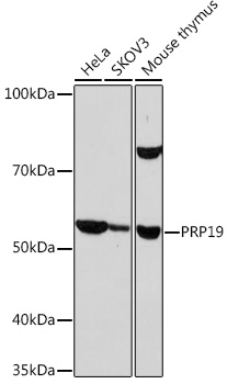
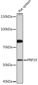
Western blot analysis of various lysates using PRP19 Rabbit mAb (A9660) at 1:1000 dilution. Secondary antibody: HRP-conjugated Goat anti-Rabbit IgG (H+L) (AS014) at 1:10000 dilution. Lysates/proteins: 25µg per lane. Blocking buffer: 3% nonfat dry milk in TBST. Detection: ECL Basic Kit (RM00020). Exposure time: 10s. |
|
|
Immunohistochemistry analysis of paraffin-embeddedMouse intestin tissue usingPRP19 Rabbit mAb(A9660) at a dilution of 1:200 (40x lens).High pressure antigen retrieval was performed with 0.01 M citrate buffer (pH 6.0) prior to IHC staining. |
|
|
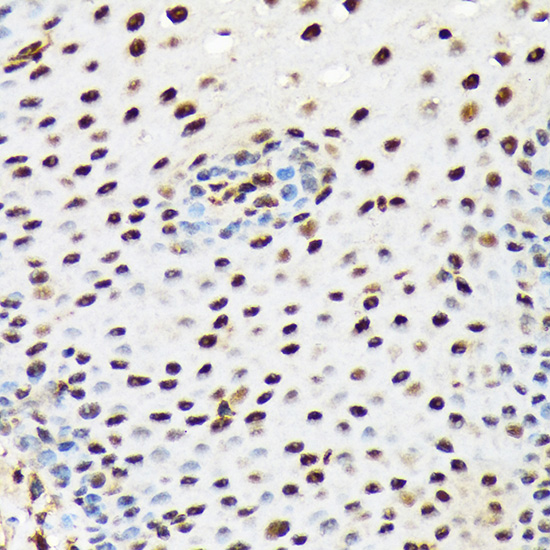
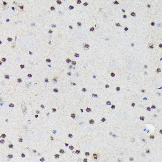
Immunohistochemistry analysis of paraffin-embeddedMouse brain tissue usingPRP19 Rabbit mAb(A9660) at a dilution of 1:200 (40x lens).High pressure antigen retrieval was performed with 0.01 M citrate buffer (pH 6.0) prior to IHC staining. |
|
|
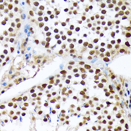
Immunohistochemistry analysis of paraffin-embeddedMouse testis tissue usingPRP19 Rabbit mAb(A9660) at a dilution of 1:200 (40x lens).High pressure antigen retrieval was performed with 0.01 M citrate buffer (pH 6.0) prior to IHC staining. |
|
|
Immunohistochemistry analysis of paraffin-embeddedRat colon tissue usingPRP19 Rabbit mAb(A9660) at a dilution of 1:200 (40x lens).High pressure antigen retrieval was performed with 0.01 M citrate buffer (pH 6.0) prior to IHC staining. |
|
|
Immunohistochemistry analysis of paraffin-embeddedHuman colon carcinoma tissue usingPRP19 Rabbit mAb(A9660) at a dilution of 1:200 (40x lens).High pressure antigen retrieval was performed with 0.01 M citrate buffer (pH 6.0) prior to IHC staining. |
|
|
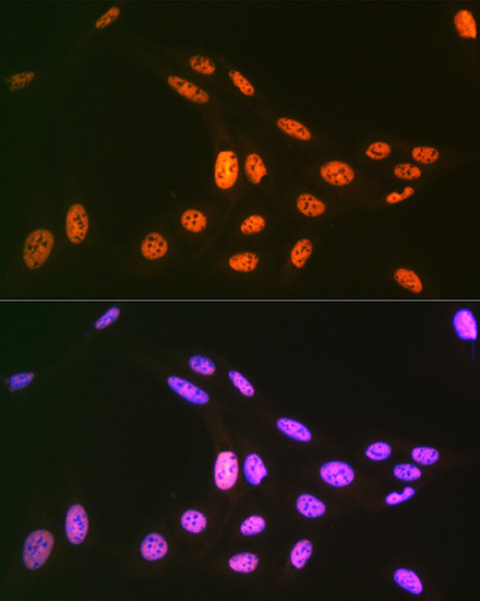
Immunofluorescence analysis of U-2 OS cells using PRP19 Rabbit mAb (A9660) at dilution of 1:100 (40x lens). Secondary antibody: Cy3-conjugated Goat anti-Rabbit IgG (H+L) (AS007) at 1:500 dilution. Blue: DAPI for nuclear staining. |
|
|
Immunofluorescence analysis of U-2 OS cells using PRP19 Rabbit mAb (A9660) at dilution of 1:100 (40x lens). Secondary antibody: Cy3-conjugated Goat anti-Rabbit IgG (H+L) (AS007) at 1:500 dilution. Blue: DAPI for nuclear staining. |
Product Guarantee and Expert Support
